Zeiss Axiovert 200 inverted microscope Room Smilow 301
Microscope uses MicroManager software ![]() or Photometrics camera software.
or Photometrics camera software.
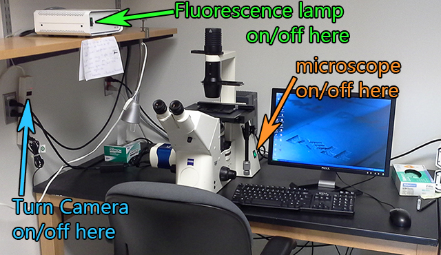
Startup Instructions 1. Turn on fluorescent light source (if needed). Shutdown Make sure you turn off the camera! |
Standard lenses on this microscope:
This camera is state-of-the-art for fluorescence microscopy. It can be used for quantification. (Technical details here.)
If you need a color camera for histology, there is an excellent upright microscope on the 5th floor of MSB. Also, the Microscopy Core and the Histopath Core have microscopes for standard color histology.
This instrument does not have any automation for changing filter cubes. However, you can set different exposure conditions for each channel and have the software save them to speed up single channel image acquisition.
Note that these lenses are not chromatically corrected, therefore, not all colors may be in the same focal plane.
This scope is good for screening and quick quantitative fluorescence snapshots of cells in dishes. There is a similar Nikon in MSB 530. For prepared slides, highly recommend using scopes optimized for this such as in MSB 530 and in Microscopy Core with Zeiss and Nikon options.
Snap images and save quickly.
You may add your snapshots (or the live window) to an Album and then save the album as a stack or output as individual files to a directory. If you have a separate album for each experimental condition, then you may take a series of pictures quickly and save them quickly with appropriate filenames. When you save an album, shoose to save as a stack of images with one name or as individual images which will have one name followed by incremental numbers.
Convert images to 8 bits all at once.
The macro for ImageJ Make_All_Images_In_Folder_8bits_v101 is useful for a folder full of tif files that may be 12, 14, 16, or 32 bits. It also may be used as a template for writing other batch processing macros with an example of getting info from the user too.
Example Images:
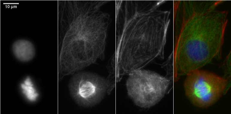
From left to right: DNA, tubulin, f-actin, merged of Molecular Probes prepared sample F14781.
40X N.A. 0.65 lens full size image of tubulin staining and the full size image that the montage was cropped from. Notice that the corners of the full field image are a little fuzzy. But the center looks good.
For a sharper 40X image, use a microscope on the 5th floor of MSB.
Example 10X image here with gamma adjustment and refocused for each color.
comments, questions, suggestions for this web page: Michael.Cammer@med.nyu.edu or mcammer@gmail.com