Zeiss AxioObserver microscope
Smilow C 17
Signup to use microscope. Must use Safari, Chrome, or Firefox; not Explorer
Instructions: [Word doc] [PDF] [Video on how to set exposure times]
This microscope is for widefield fluorescence, simple brightfield, Nomarski/DIC, crossed polarization (see below), and phase contrast. It has automated focus for Z series and precision stage for tiling large areas or repeatably imaging multiple positions. It may be used for live imaging and for fixed samples.
A few of the specifications:
- Zeiss AxioObserver.Z1
- 5X lens
- EC Plan-Neofluar 10x/0.30 Ph1 WD=5.2
- Plan-Apochromat 20x/0.8 WD=0.55 M27
- EC Plan-Neofluar 40x/0.9 Pol WD=0.41 M27
- EC Plan-Neofluar 40x/1.3 WD=0.21 M27
- Plan-Apochromat 63x/1.40 Oil DIC M27
- DIC Sliders for /20x, 40x and 63x
- Cond LD 0.55 H/DIC/Ph 6x Mot
- FL Filter Set 49 DAPI
- FL Filter Set 38 HE GFP
- FL Filter Set 43 HE Cy3
- FL Filter Set 50 Cy5, EX BP640/30 S free
- FL Filter Set 46 HE YFP shift free available on request
- FL Filter Set 47 HE CFP shift free available on request
- 50/50 mirror available on request
- Light Guide HXP120, 2m
- Axiocam 503 Monochrome camera
- Axiocam 503 color camera
There is no filter set for Alexa Fluor 594 but the filter set for Cy3 works well.
People often ask, what about 100X? The 63X N.A. 1.40 lens has the same spatial resolution at the 100X lens when matched with the small pixel AxioCam on this microscope but with benefits such as a wider field of view. Also, a magnification lens my be placed in the lightpath for spatial oversampling.
Example Images. Click on images for larger versions.
All the micrographs in this figure were taken with the AxioObserver: https://www.ncbi.nlm.nih.gov/pmc/articles/PMC9720609/figure/F3/
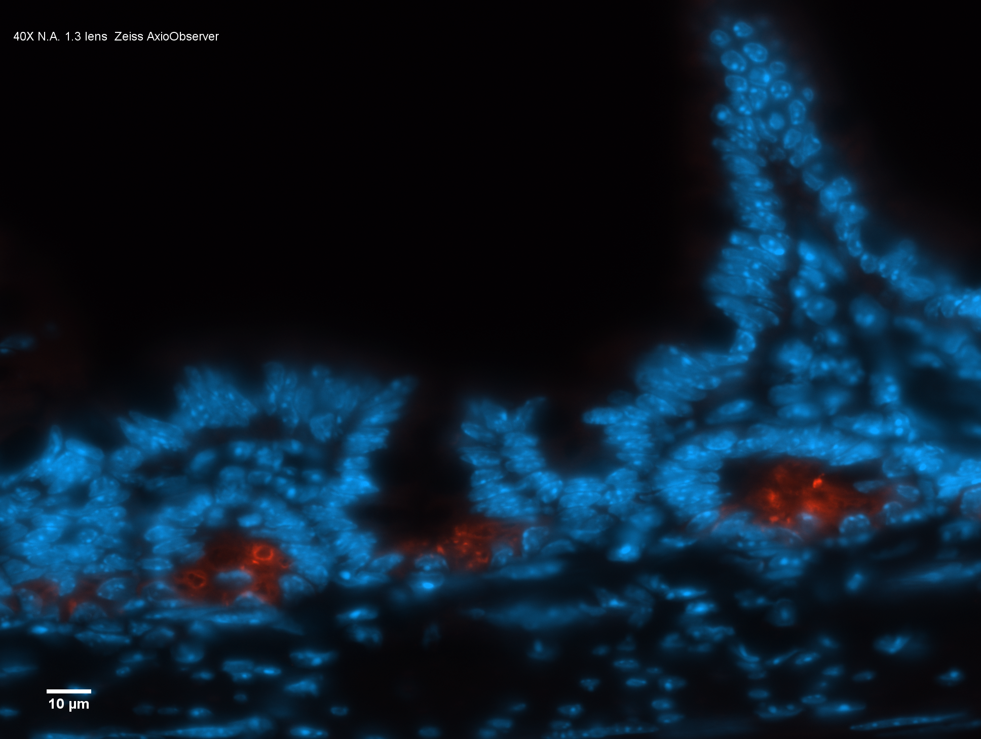
Example fluorescence imaged with 40X NA 1.3 lens. For full resolution, click on image.
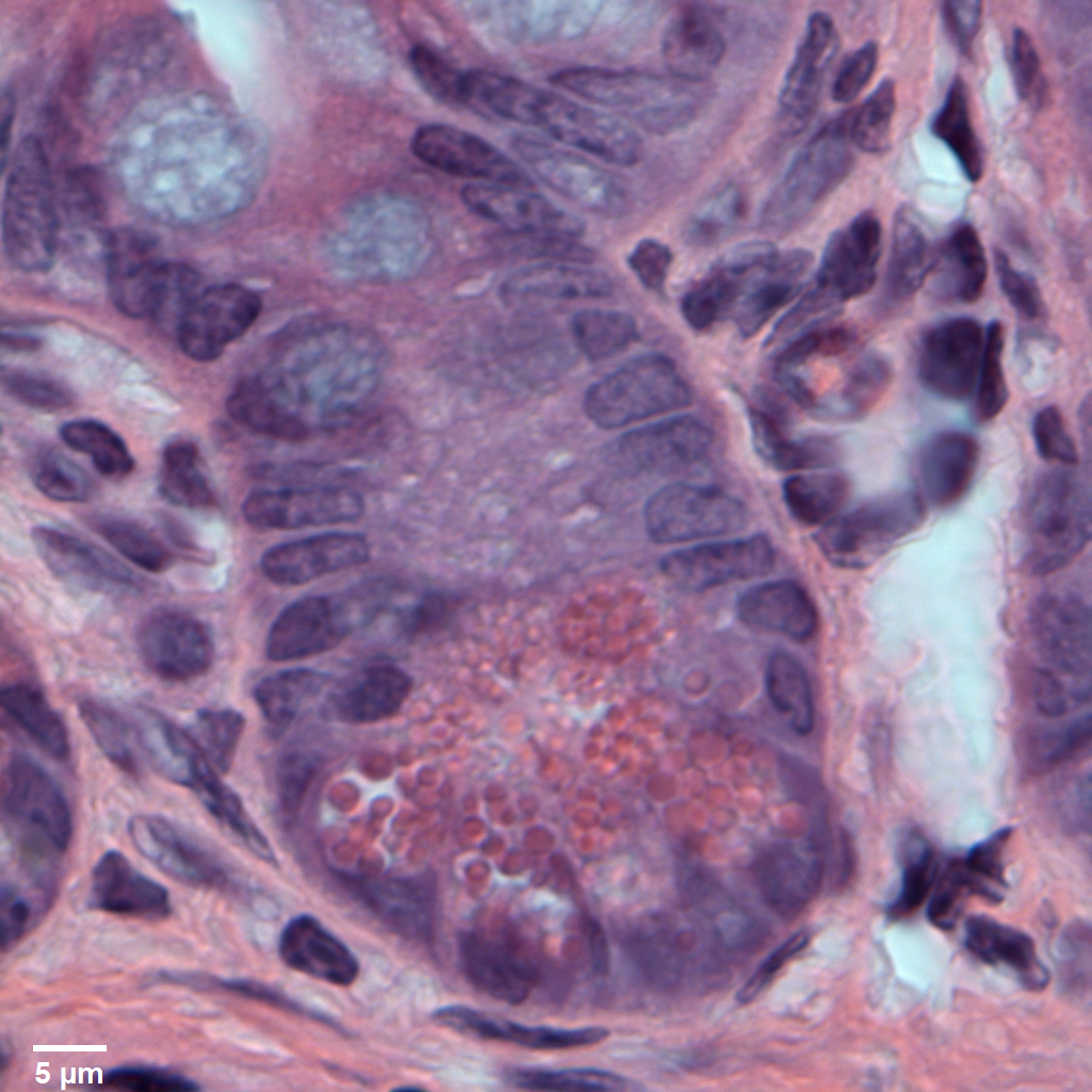
Example H&E at highest magnification. Micrographs often not crisp because tissue sections are thicker than the depth of field of the lens.
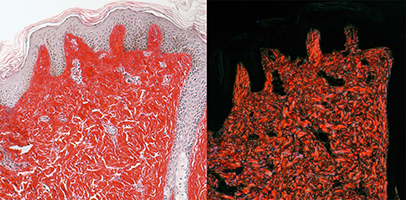
Example of crossed polarization showing collagen,click on image for web page with explanation.
Old website here for reference: index_prejune2018.html
SN 3851000366