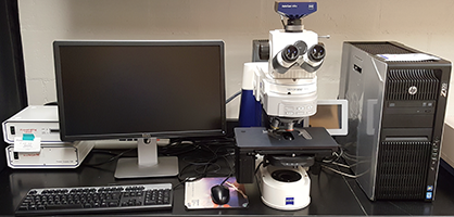
Zeiss AxioImager.M1
MSB 530
(last updated 20160622_0844) |
 |
To sign up in advance:
http://signups.med.nyu.edu/facilities/equipment/signups/pathology-zeiss-axioimager
To Turn on :
- If using fluorescence, lamp power supply labeled "HBO 100"
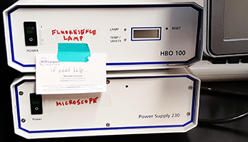
- For microscope operation, turn on Power Supply 230 and wait for touch screen at right to show system initialized before running software.
- Computer on.
- After microscope fully turned on, software AxioVision Rel. 4.8

To Turn off :
- Transfer all files. We recommend this.
- Quit AxioVision.
- Check if anyone is coming in after you.
If yes, leave on.
If not, then continue shutdown:
- Turn off microscope power supply.
- Turn off mercury arc lamp.
- Shut down computer. Yes, please turn it off.
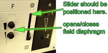
If you are shooting brightfield, make sure you Köhler align:
If imaging by brightfield, phase contrast, or DIC/Nomarski, always set condenser for Kohler illumination.
1. Focus on sample
2. set condenser for BF
3. Close Field Diaphagm
4. Move condenser up and down using knob on right until center octagon is in focus
5. Center by turning two knobs on condenser.
6. Open field diaphragm to edge of visible field, not all the way.
More explanation of Köhler illumination here.
Tips:
- If imaging brightfield, set up Kohler illumination.
In addition to the instructions for Kohler, make sure the condenser iris setting is equal to or greater than the NA of the lens.
- "Microscope Control" button for fluorescent filter & shutter control.
- Manually turn objective turret slowly to change lenses.
- Right click on Live window to get Properties window.
- In properties, use tabs at top of window.
- "6D-Acquisition" button to get window for setting up multicolor snaps or mosaic imaging.
- In the Channels tab, right click on small colored boxes in row at top to turn on and off for automated acquisition.
- For each channel find the exposure in live mode and then click fixed exposure button and type in. This is critical if making intensity measurements. Set exposure for each channel based on brightest condition and do not change.
- Always save images in native Zeiss ZVI format or as TIF. Do not save as JPG because it is lossy.
YOU NEED TO KEEP TRACK OF THE MAGNIFICATION. Do at least one of the following:
- Put the magnification, such as "10X" and "40X", in the folder names and sort images into the correct folders.
- Put the magnification in the name of every file you save. E.g. "WT Hek G tub R actin 20X"
- Use only one magnification for all of the images and keep good notes.
- Later you may look here for spatial scales for scalebars and measuring distances.
- If you save as TIF and want to save raw data for quantification of intensities, make sure you unclick the boxes as pictured. Setting to convert to 8 bits and/or apply mappings will lose the raw data.
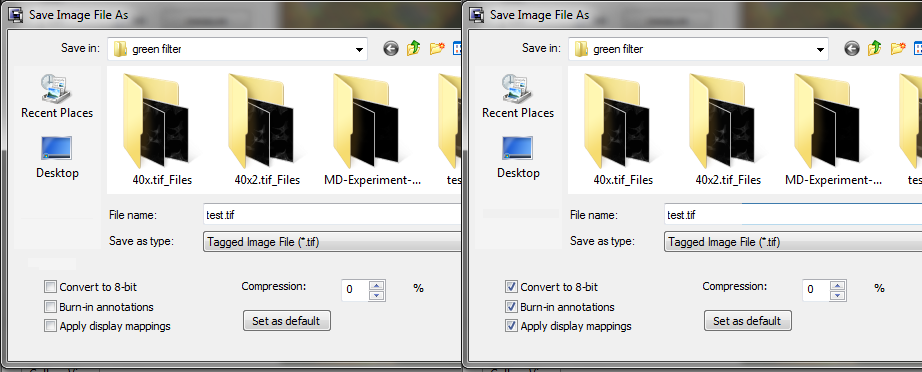
At the left, the checkboxes are unchecked to save the raw data.
At the right, the raw data are not saved.
- Thinner samples (coverslipped slides) need to be put on spacer on microscope stage to reach 63X lens.
Simply stack your slide on top of a blank extra slide.

- Warning: Using the 63X lens you may not get full resolution images because the microscope is not on an anti-vibration table.
Example movies of the problem here or here.
About the instrument:
Lenses:
5X NA 0.15 EC Plan-Neofluar Spatial Scale
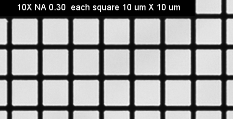
10X NA 0.30 EC Plan-Neofluar 420340-9900 Spatial Scale
20X NA 0.50 EC Plan-Neofluar 420350-9900 Spatial Scale
40X NA 0.75 EC Plan-Neofluar 420360-9900 Spatial Scale
63X NA 1.25 EC Plan-Neofluar 420480-9900 (oil) Spatial Scale
For best images use #1.5 coverslips. The lenses are designed specifically for #1.5 coverslips. Says so right on the side of them.
Camera: AxioCam MRm Rev3, 12 bits grayscale
This means that the intensity range is from 0 to 4095.
Filters:
Green (GFP, Alexa488, etc. ex450-490 FT510 BP515-565
Red (Cy3, RFP, etc) BP560/40 FT585 BP630/75
Blue (DAPI etc):
G365 FT395 BP445/50
near infra-red (Cy5, Alexa647, etc) [click here]
Software: AxioVision 4.8.2.0
Only installed multidimensional acquire modules are:
channels
mosaic (tiling)
(Z stack and other software modules not installed.)
Computer: 32 bit Windows 7 with 16 GB RAM
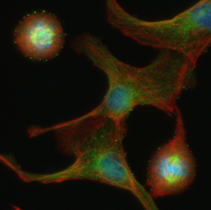
Example image of f-actin (red), tubulin (green), and DNA (blue) using the 40X N.A. 0.75 lens. Nuclei are approximately 10 um diameter.
Click here for full field view linear scaled raw data.
Click here for full field view local contrast enhanced.
<-- Back
comments, questions, suggestions for this web page: Michael.Cammer@med.nyu.edu or mcammer@gmail.com