a few results from VT-iSIM demonstration on site
Februrary 2016
Microscope: http://www.visitech.co.uk/vt-isim.html and http://www.biovis.com/vtisim.html
live worm (6.5 MB)
nuclei (12 MB)
HEK MTs (7.5 MB)
neurons (15.5 MB)
dendritic cells (4 MB)
mitochondria (10+ MB)
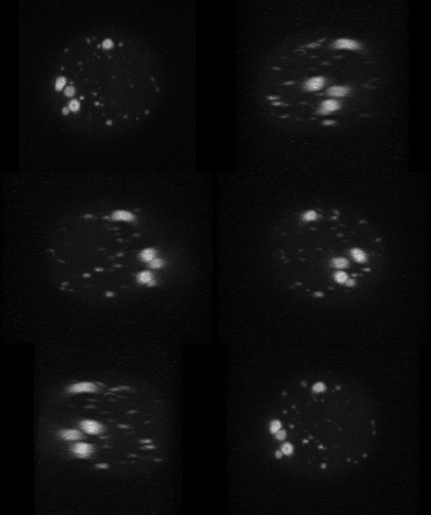
The following is an example of an approximately spherical live cell in culture that has fluorescent protein containing organelles (like vesicles) moving inside. These are rare occurances so more than twenty fields were imaged. Each field had a Z series collected. This produced approximately 1 TB of data per night. Then the few events during the overnight run were extracted making each relevant dataset a few GB.

