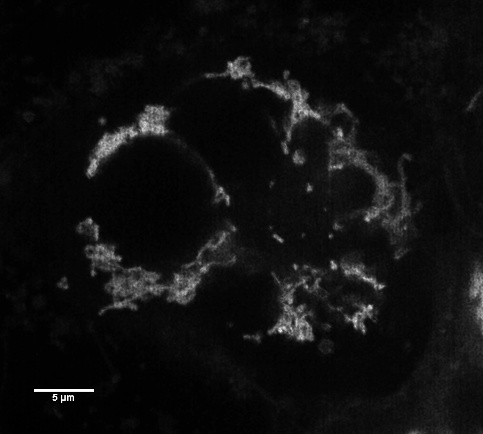
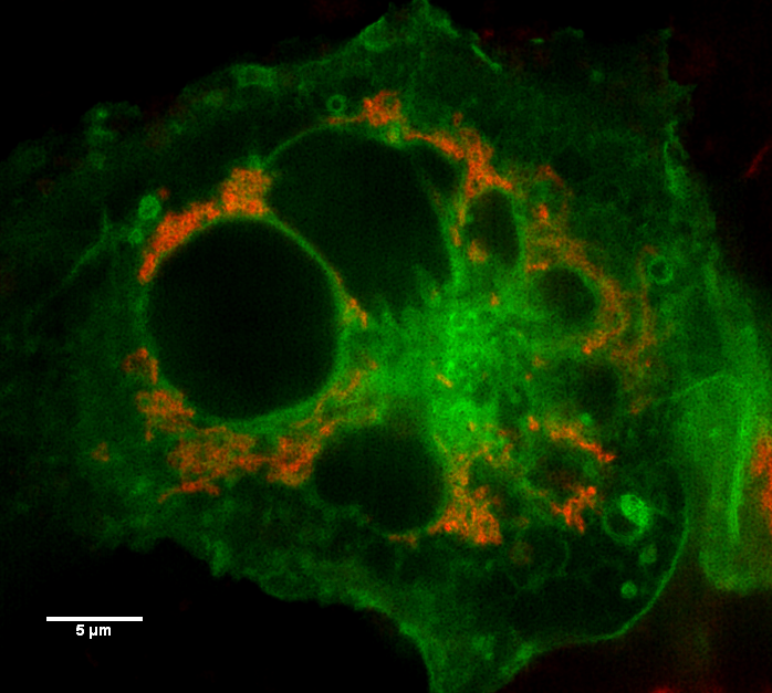
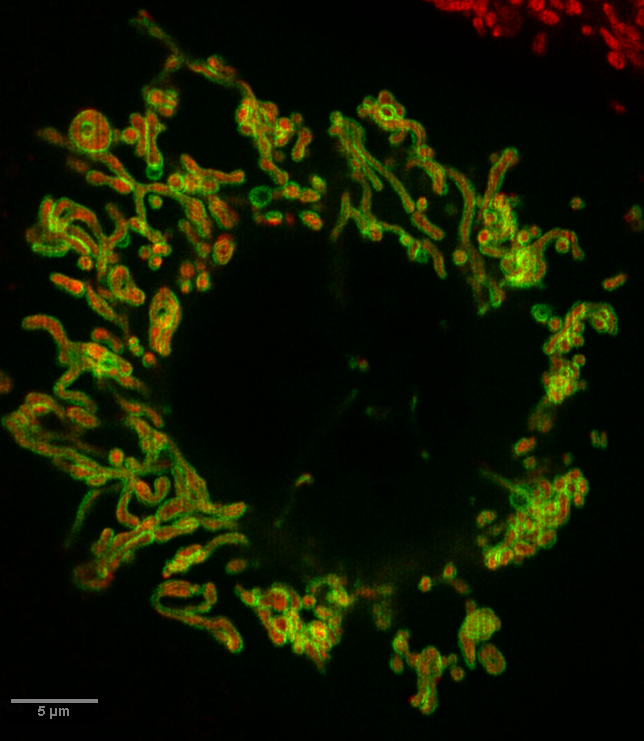
mitochondria red
other GFP-fusions green (not identified here)
VT-iSIM demo Feb 2016
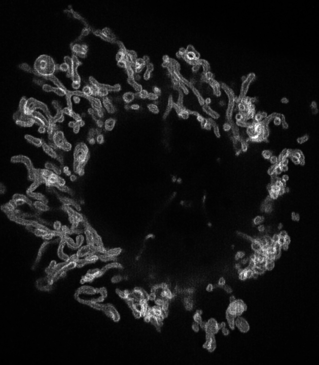
From Z series, deconvolved, median filter of slices 20-24.
Original spatial scale from microscope.
The Z series (at 67% spatial scaling).
also available here as AVI with jpg compression
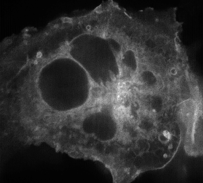
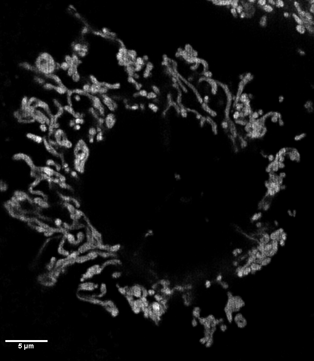
Maximum pixel projection of planes 13-15 with fft mask applied to remove vertical lines.
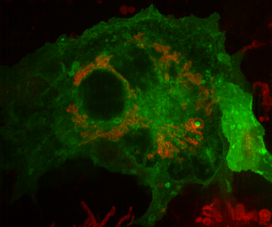
Another example, maximum pixel projection of slices 15-17 of Z stack taken at 0.2 um steps. XY pixel size reported as 0.057 X 0.057 um (oversampled -- in theory best resolution 100 to 125 nm, not 57 nm). Red green notin perfect register because cell moving fast and colors have to be taken in sequence; this is one possible limitation of the instrument.
The green is really on the outside of the red mitochondria but this can only be resolved along the sides, not when the green is a thin sheet in the plane of focus above & below.
also available here as AVI with jpg compression