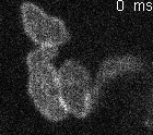

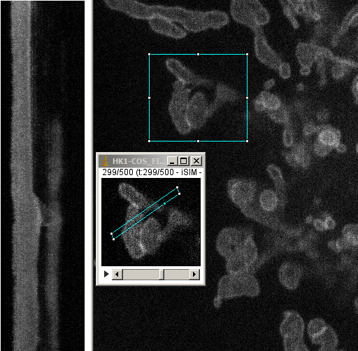
imaging GFP fusion with protein localized on the surface of mitochondria in COS cells in culture at 37 degrees C.
VT-iSIM demo Feb 2016
Single plane time series at 100 ms exposures with no pause between snaps, a.k.a. 10 fps.
Each pixel is either 0.065 um or 0.1 um linear.
|
|
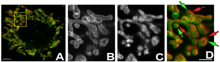 |
| Requirement for dual cameras for simultaneous two channel imaging of live cells. Two fluorescent channels out of register with serial exposures as shown in this deconvolved single optical section of live COS cells expressing HK1-GFP and loaded with mitotracker dye. Protein localization on outer mitochondria membrane surface (B, green) clearly resolved from interior (C, red). Arrows point to mis-registered pixels due to movement between exposures. (A) Field at 100X, scale bar 5um. (B) HK1-GFP; (C) Mitotracker; (D) Merged B and C, scale bar for B, C and D, 2um. |