There are many 3D software options, some free and some expensive, and the cost doesn't necessarily correlate with the features.
The simplest one, the one we go to the most, is in the ImageJ Image > Stacks menu.
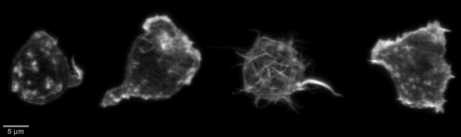
The following four cells are fom a single field collected with a Yokogawa spinning disk in 2012. Resting t cells plated on a coverslip were fixed and stained with Alexa 488 phalloidin (prep by Dr. A. Zanin-Zhorov in Dr. ML Dustin lab) and imaged with an Olympus 60X TIRF lens on an IX70 with spinning disk and a Hamamatsu Orca ER camera operated by MicroManager and originally assembled by Solamere. Only contrast enhancement was linear in the 12 bit to 8 bit conversion. Each cell was cropped from the field, 3D projected with ImageJ, and Image > Stacks > Tools > Combine... to make the four panel figure below. (This last step may be automated.)
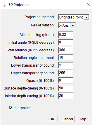
This is a simple macro to make a 3D rotation of a confocal dataset. The first block does the rotation. The second part prepares it for export as a movie for presentation.
// User needs to draw a box over cell(s) first or entire field will be processed.
// To rock instead of 360, change initial=330 total=60.
// To rotate on X instead of Y axis, change to axis=X.

	t = replace(getTitle, ".czi", "");
	t = replace(t, ".tif", "");
	getVoxelSize(xwidth, yheight, depth, unit);
	run("Duplicate...", "duplicate channels=1-2");
	run("3D Project...", "projection=[Brightest Point] axis=Y-Axis slice=0.50 initial=0 total=360 rotation=5 lower=1 upper=255 opacity=0 surface=50 interior=35 interpolate");
	rename(t + "_3D");
	setVoxelSize(xwidth, yheight, xwidth, unit);

	run("RGB Color", "frames keep");	// The original macro recorder said "slices" instead of "frames".
	run("Scale...", "x=3 y=3 z=1.0 width=372 height=339 depth=33 interpolation=Bilinear average process create");
	run("Scale Bar...", "width=5 height=5 thickness=2 font=11 bold label");
	rename(getTitle+"_RGB");
Click for a few examples of this method used on tissue samples imaged by multiphoton.
As of 20190318, here are a few other 3D reconstruction software we have used recently. This list is by no means exhaustive.
My all time favorite was VoxelView from the early 1990s on a SGI computer.