Zeiss AxioZoom microscope
Smilow C-17
This is a macro- to microscope, a.k.a. a dissection microscope, for looking at transparent objects on the cm scale to cells on the 25 um (or so) scale. Software includes tiling and focusing through a large depth.
It has a monochrome camera for fluorescence and a color camera for brightfield and relfectance imaging. In some cases, you may want to use the color camera for fluorescence, but in most cases, the monochrome camera is for fluorescence.
Standard filters for fluorescence are blue, green, red. Others may be available by request.
To use microscope you must be trained by Michael Cammer or Yan Deng via the Microscopy Core.
** Signup to use microscope **
There is also
a dual goosneck light source.
https://www.amazon.com/AmScope-LED-6W-Powerful-Gooseneck-Illuminator/dp/B007LBELH4/?th=1
One of the nice things about LED instead of halogen is that the light does not heat the sample.
You may also find the LED desk lamp in the room useful for additional ambient lighting.
Starting up
- Start up computer and log in (password taped to the screen).
Go to iLab and start your Kiosk session.
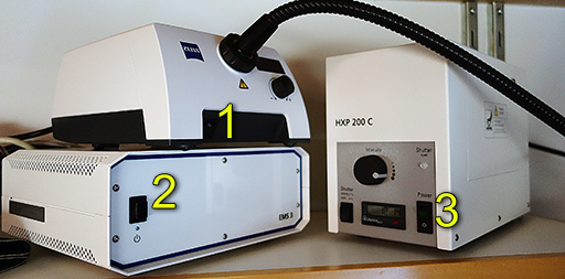
Example pictures here and here. - Optional: Switch 1 is transmitted light. Use adjustment knob to make brighter or dimmer. You only have to turn this on if you want transmitted light from under the sample.
- Switch 2 is microscope controller. This is required.
- Optional: Switch 3, is for fluorescence. Use adjustment knob to make brighter or dimmer.
- After the touch screen is on, you may run Zen software.
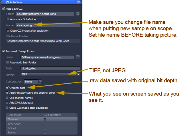
All fluorescence images should be saved in czi format.
Transmitted or brightfield images may be saved as 8 bit RGB tif for convenience, but they will not have metadata for spatial scale and the contrast is non-linear and arbitrary. Therefore, we recommend setting autosave and export on together to save in both formats. Details below.
When using the computer, only store files on Drive D:
DO NOT STORE FILES ON DESKTOP or anywhere on Drive C: including "Pictures" folder.
Shutdown
- Transfer files to a server such as \\research-cifs.nyumc.org\research.
DO NOT plug a USB device into the computer. This is forbidden.
- Quit Zen.
- MAKE SURE LENS IS SET TO LOWER MAGNIFICATION AND FOCUS IS RAISED BEFORE TURNING OFF CONTROLLER BOX!!
- Upper right touchpad, use shutdown button to turn off box 2.
Wait for this to finish.
- Check whether someone is coming in soon after you before turning off the lamp for fluorescence.
- Switches 1 & 3 off.
- End iLab Kiosk session.
- Shut down computer.
- CLEAN UP!
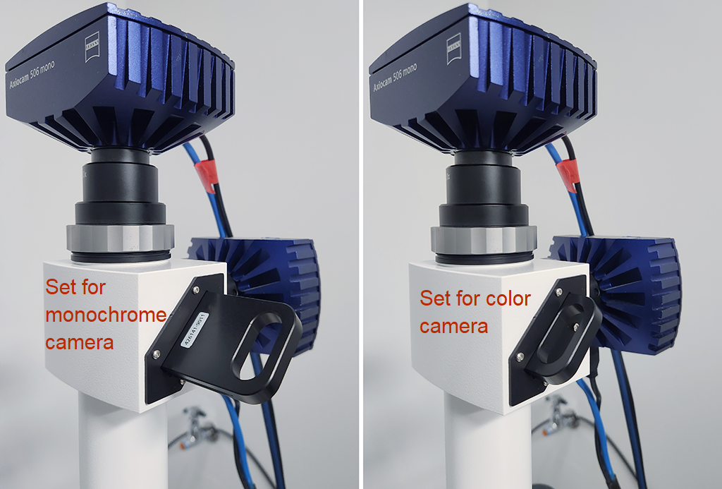
There are two cameras on the system.
Use the monochrome camera (on top) for fluorescence imaging.
Use the camera at the back for color brightfield or reflectance imaging.
To switch between the cameras, there is a manual slider.
Push in all the way for the color camera.
Pull out one click for the monochrome camera.
DO NOT REMOVE the mirror from the camera splitter!!
If you want to see the Zeiss technical manual, it is here.
Protocol for 96 well plates here. Warning: This may have changed in August 2026.
About the microscope
| Button |
Useful for... |
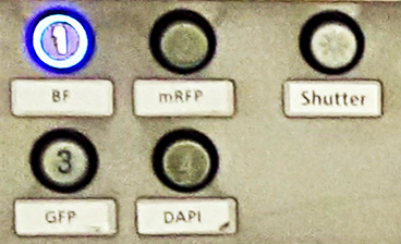 |
| Dapi |
Dapi, CFP (maybe), Pacific Blue
Zeiss set 49: G365 - FT395 - BP445/50
|
| GFP |
FITC, Alexa488, GFP, YFP
BP470/40 - FT495 - BP525/50
|
| mRFP |
RFP, mCherry, rhodamine, Cy3, Alexa568, Alexa594, propidium iodide
BP572/25 - FT590 - BP 629/62
|
| BF |
Transmitted light. (Also reflected light with an additional light source; sometimes there is one in the room or you could bring your own.) |
| Shutter |
Light on/off |
| |
The filter cubes in this system are standard Zeiss and are simple to snap in and out. This means that if you have special spectral needs, we can swap out the blocks easily for your specific needs. We just need the block... |
|
| |
Crossed polarization methods are available and examples. |
|
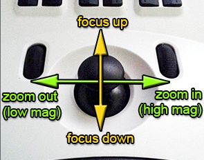 |
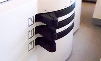
Push down to toggle to stage moving mode.
Push down to return to focus/zoom. |
When imaging fluorescence, to minimize reflections and maximize contrast, recommend these base settings.
You want to make sure the space below the sample is as blackand non-reflective as possible. (More details here if interested.)
For transmitted light imaging, recommend playing until you have the ideal image and then set the same every time for repeatability.
Lenses:
There are two lenses.
To change from one lens to the other, simply grap the lens and rotate to the next position.
The higher magnification lens focuses lower and closer to the sample. When switching to this lens, a message will be displayed on the touch pad. Please read it and follow thie instructions so that the lens does not crash into your sample or the stage.
Software Startup:
Warning: This section needs to be updated based on new software installed in August 2026.
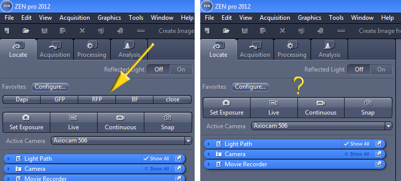
On software startup the defaults should have settings in the Locate tab for all fluorescent colors and brightfield.
| This is how the Locate window should look. The yellow arrow points to a row of buttons that control the microscope. |
If the buttons are missing, then you need to reassign them using the Configure... button.
We are trying to figure out why the buttons sometimes disappear so that we may stop this from happening.
Please let us know if you you know how to fix this. |
 |
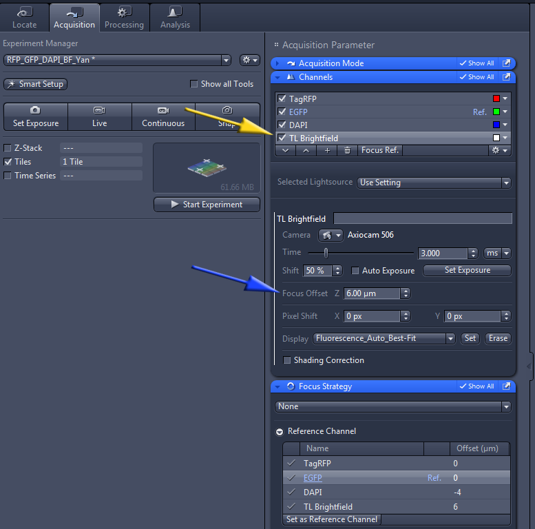
To preview and set up acquisition parameters for each channel, in the Acquisitiion tab click on the channel (yellow arrow below) and Show All in the blue bar above it. Note that you may set different focus settings for different channels.
Software installed May 2016 should have this Auto Save menu option for saving files:
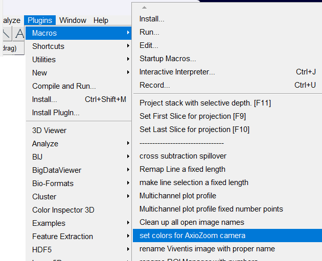
When importing color camera CZI files into ImageJ/Fiji, you will need to use the macro "set colors for AxioZoom camera" bundled in MC_macros.ijm. Set view stack to Hyperstack and color mode to Composite.
You may then need to set the contrast for each channel usingthe Maximum slider in the Brightness/Contrast window. If you have multiple images to open, you can set the contrast for one of them and using the Set button at the bottom of the Brightness/Contrast window, propagate the settings to all the open images, and save them as OME TIFF.
Crossed polarization methods are available and examples.
<-- Back
comments, questions, suggestions for this web page: Michael.Cammer@med.nyu.edu or mcammer@gmail.com