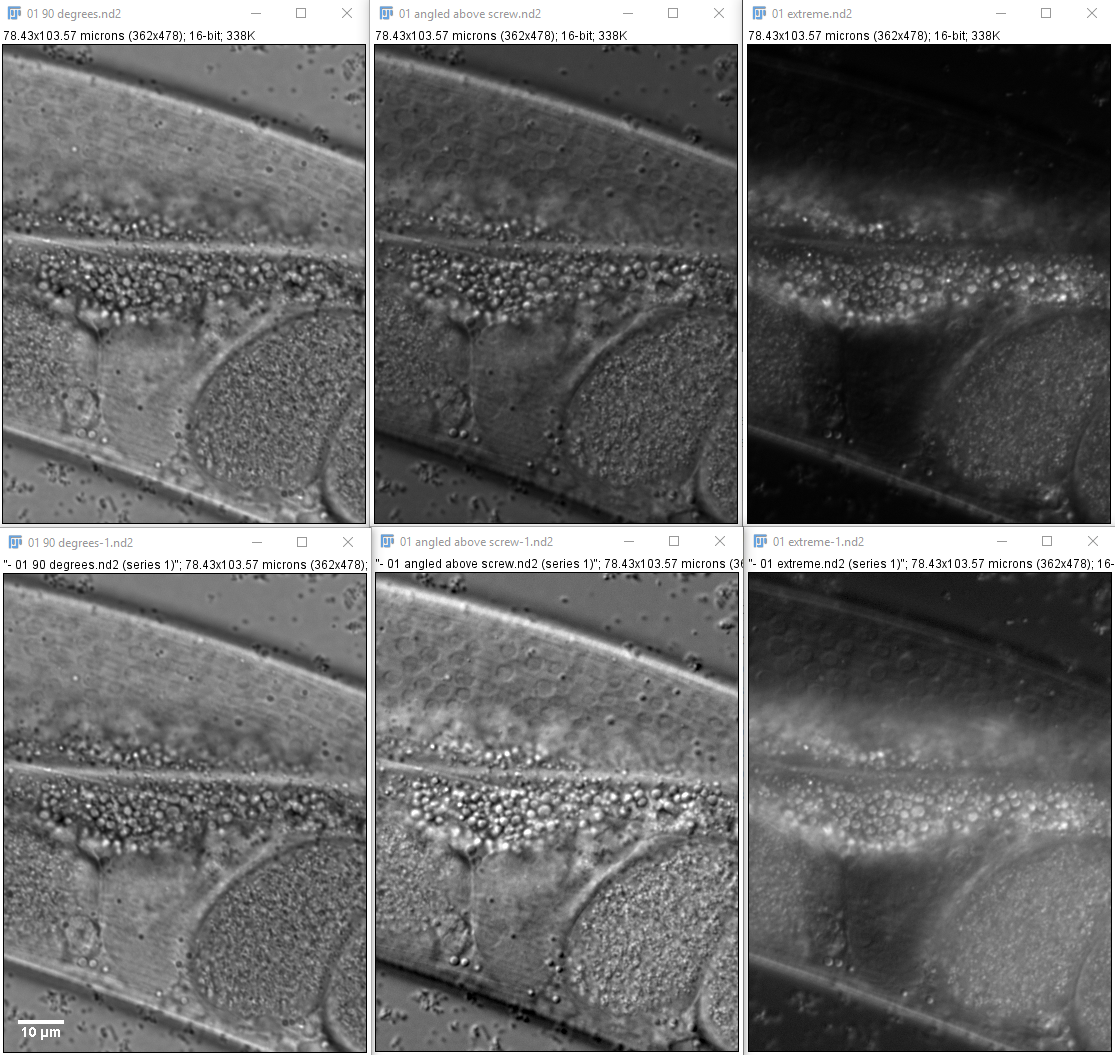
The top row is with contrast from min to max values (essentially raw data linear) and the bottom row adjusted with a gamma curves the way I think the pictures look good.
Sample is a live worm.
Nomarski / DIC on Nikon Spinning Disk microscope
last updated 20200920 1615
Recommend order of experiment Lambda Z because of slow speed of CSU in/out.
May also image DIC through the spinning disk.
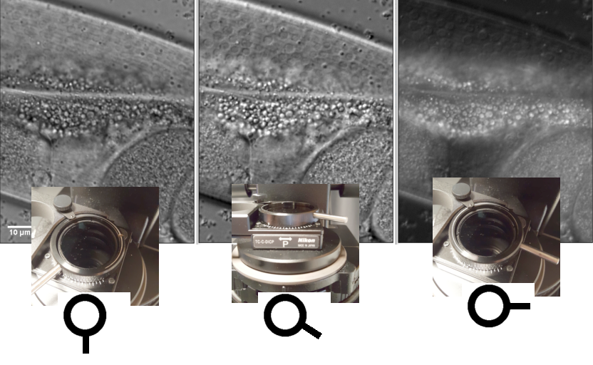
From left to right, least extreme to most extreme Nomarski in one of the directions the optics may be adjusted.
The top row is with contrast from min to max values (essentially raw data linear) and the bottom row adjusted with a gamma curves the way I think the pictures look good.
Sample is a live worm.
The polarizer on the condenser may rotate in a range of 180 degrees to adjust the magnitude and apparent direction of the the contrast. To the right is most extreme crossed polarization. These pictures show rotations towards the front of the microscope but this is symmetrical to the rear of the scope too.
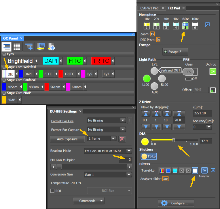
To make an Optical Configuration button for this, you can replace the brightfield button or copy the brightfield button to a new one and switch from a Turrt-Lo blank position to position 6 which will automatically set Analyzer In, then lock in the new setting with the little traingle to the right of the OC button.
You may use the Widefield buttons to have the spinning disk move out of position or add an OC to the Confocal row of OCs to image through the spinning disk unit; this requires a longer exposure time, but is faster due to harware not moving.
When using Twin Cam, DIC will be collected by both cameras. If you want to collect a DIC and bright fluorescent image simultaneously, we can do this by putting a bandpass filter on top of the polarizing filter in the condenser. (We haven't tried this yet, but it should work.)
Beware:
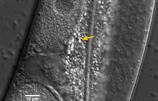
Smudging or bleeding looks bad and may damage the camera.
To avoid it:
Keep exposure times 1 frame or longer.
(Approx 34 ms at 30 MHz readout and 112 ms at 10 MHz readout.)
10 MHz may prevent smearing.
DIA light set 50% or less.
Maximum pixel value less than 20000 (only need 4000 to 6000 for a good image).
EM gain low (3 to 10).
We need to figure out how to deal with the following gain issue:
If switching gain from one exposure to another, the images with have unacceptable shading from top to bottom.
Therefore, we may need to keep gain high to match fluorescence and be very careful with the diascopic intensity not to burn the camera. Or insert a dummy image collection before and after the transmitted image to reset the gain and delete the images at te end.
(Other labs do this for widely varying fluorescence signals.)
For Lambda Z methods, only the first image of each Z series will have the shading, so can easily solve by adding additional plane at beginning, but for Z Lambda or single planes with quick channel switching, this is a problem.