From left to right, set at 8, 10, 12, 14.
Some people may prefer 8, some 10, and you can imagine what 9 would look like.
airyscan_20171206
The following is AiryScan imaging of gamma tubulin staining in Drosphila testis. This sample was not intended for AiryScan, but we needed a sample for testing, so tried it.
We did not expect we would resolve the centrioles to the extent that we could see hollow columns, but it worked.
The deconvolution setting is empirical and may be to taste.
From left to right, set at 8, 10, 12, 14.
Some people may prefer 8, some 10, and you can imagine what 9 would look like.
Enlarged for easier viewing.
More examples:
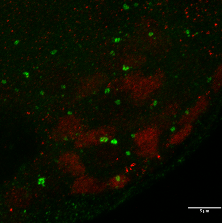
A maximum pixel projection of the AiryScan deconvolved stack with setting at 10 to show the overall morphology of the tissue. Scaled 50% and cropped.