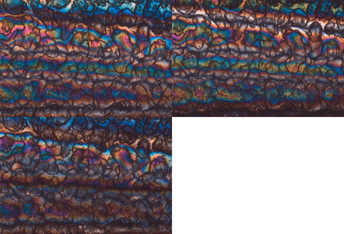
Polarized light images are acquired with the AxioZoom by placing the sample between two crossed polarizing filters and taking three pictures, careful to turn the paired plane polarized filters between the exposures without moving the sample.
The images are threfore at 0, 30, and 60 degrees to repesent a full 360 degrees rotation. Effectively, 0 and 90 degrees give the same result in this system. (You may make your own template for 15 or other degree rotations, but 30 degrees should be sufficient.)
The rotations are important because, especially in biology, we do not have control over the orientations of the structures we need to image and these structures may also turn, which would require different angles to capture them.
This animation cycles through the three angles of rotations of the light. The material is the handle of a clear extruded plastic knife.
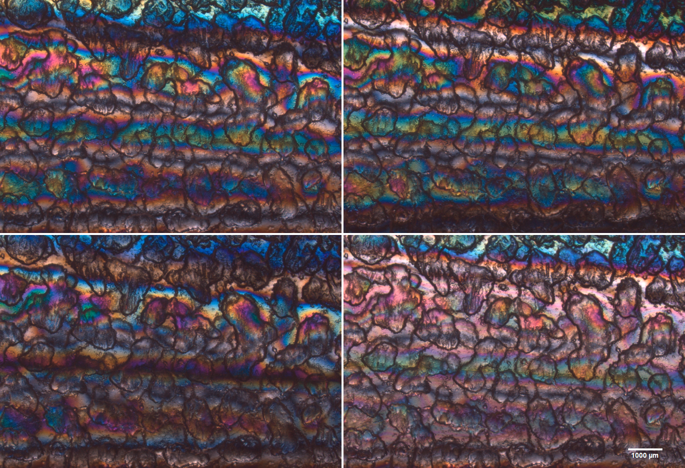
This montage shows the three rotations each static and the bottom right image is the maximum intensity projection of the three raw data images:
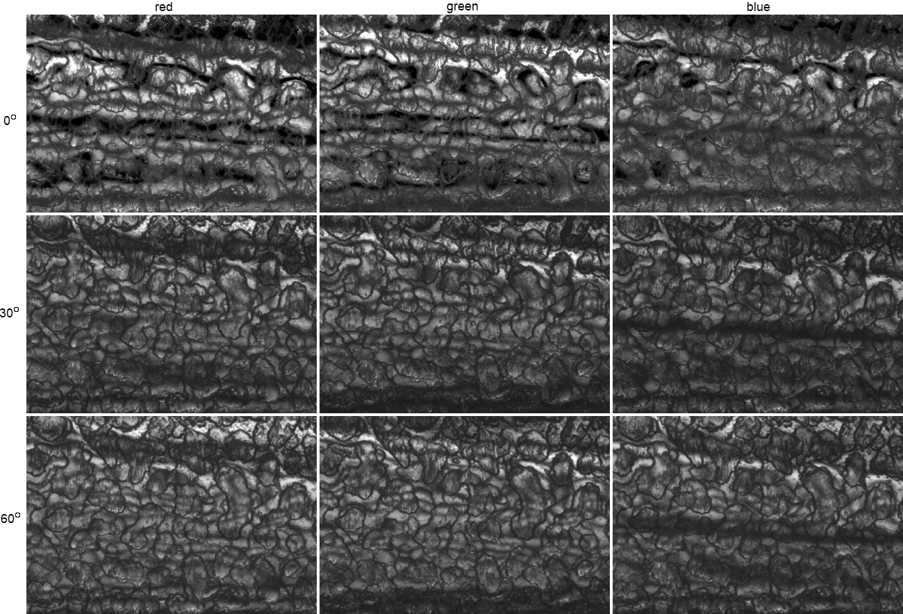
The following montage shows each row one of the angles and each column a different color separation. The images were collected with a RGB Zeiss color CCD camera.
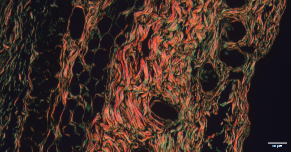
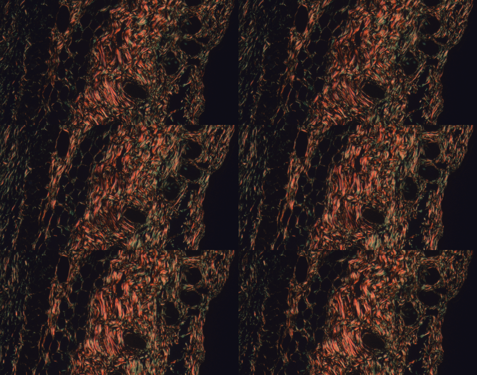
Most users of the AxioZoom will be imaging biological material. The following example is of sectioned skin stained with picrosirius red.
Note that the callagen bundles appear continuous in this maximum intensity projection of a series of plane polarized light images taken at different angles. The animation below, of the individual images, makes the bundles appear discontinuous. This may be a metric when comparing biological conditions.
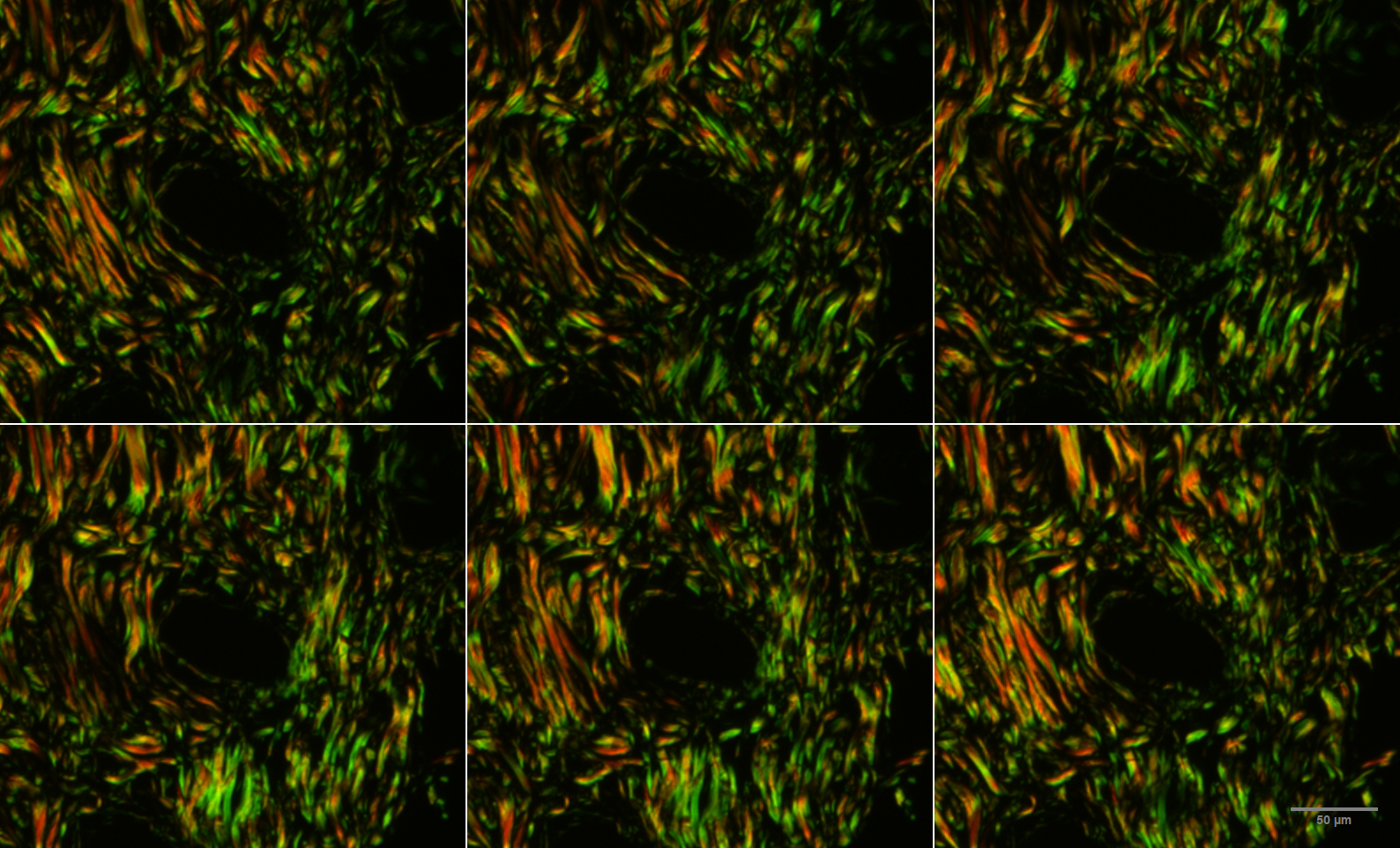
There is a lot more flexiility of image analysis and image processing when the 14 bit CZI files are used instead of the 8 bit TIF version above. The following images were taken at 15 degrees intervals. These are from the CZI format images:
Instructions for saving files
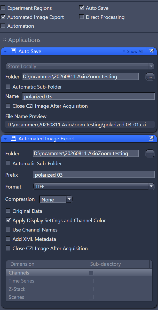 |
Each time a new rotation series (group of three images) will be collected, make a new file name. This will make grouping the files easier. The files are saved as both czi and as tiff. The CZI data may be used to quantify each channel. This format also saves the magnification and other instrument parameters. The TIFF format saves what you see on the screen. This means that contrast settings, including gamma, are applied to the image. This is very convenient for showing the images later, but may not be useful for quantification.
When you change the file name in the Auto Save window, it propagates to the Exorrt window. But if you type it in the Export window, it does not update in the Save window.
When importing color CZI files into ImageJ/Fiji, you will need to use the macro "set colors for AxioZoom camera" bundled in MC_macros.ijm. Set view stack to Hyperstack and color mode to Composite.
|
When you open the images with ImageJ, the TIFs may be assembled using Image > Stacks > Images to Stack.
The CZI files may be assembled using Image > Stacks > Tools > Concatenate...
The original data, which may have am intensity range from 0 to 16383, has a lot more leeway for contrast adjustments and quantification.
Click here to see the original design for this.