20160310 1830 to 1920
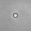
4 um diameter Tetraspeck bead embedded in 1% agarose in capillary with black band
imaged in Sigma Histodenz prepared by Yan Liu
20X CLARITY lens
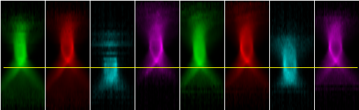
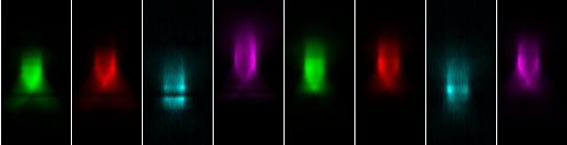
Progressive Z slices at 0.9 um steps through 4 um Tetraspeck bead.
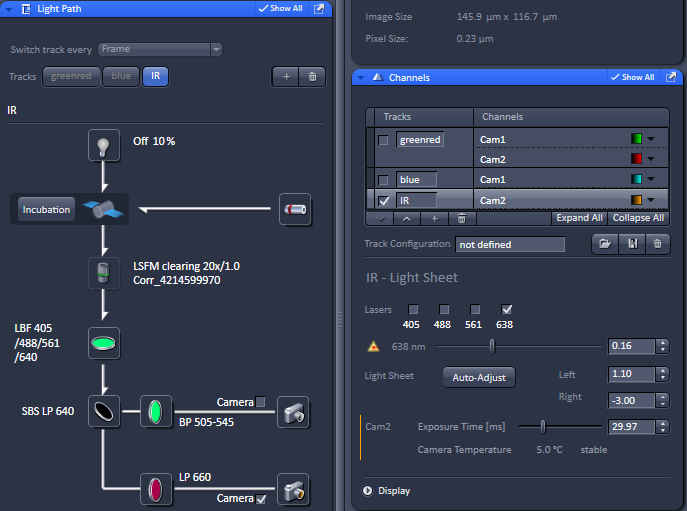
Channels from top to bottom: green, red, blue, far-red.
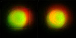
Yellow lines are guides on X axis showing camera 1 shifted to left of camera 2.
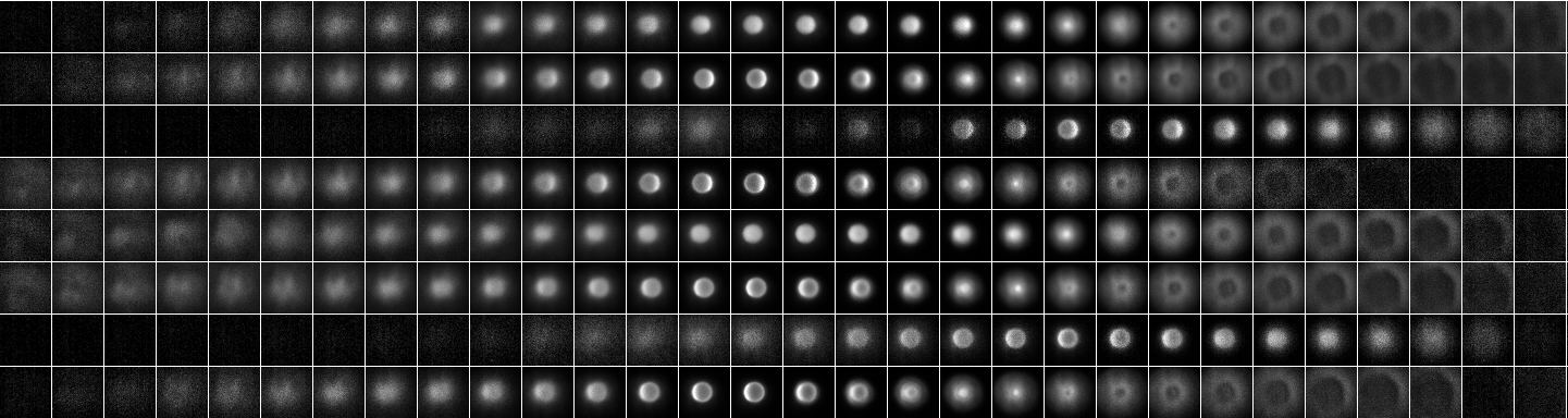
Z series montage, this one with filters changing at each Z slice ("Frame").
Top 4 rows left illumination.
Bottom 4 rows right illumination.
Channels from top: G, R, B, IR, repeated.
Left to right Z slices.
Contrast adjusted per slice.
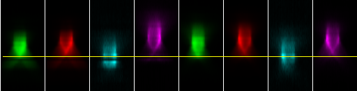
This XZ reslice shows that filters were turning or laser not on properly during one of the blue channel collections.
Blue channel focused low probably due to chromatic aberration.
The horizontal yellow line is a guide to better see Z shifting of different wavelengths.
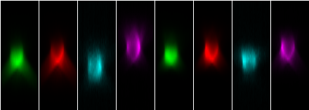
Another Z series with each color images at each Z. This is slow and we expected to give better results than Z of each frame, but again odd blanking and bright bands in blue (405 nm excitation) channel and chromatic aberration worse.
Full Z series of each channel.
These XZ reslices show much more consistent laser instensity through each Z series and was very fast to image.
But chromatic aberration appears worse.
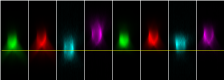
Can the appearance of chromatic aberration be corrected by changing the lightsheet position?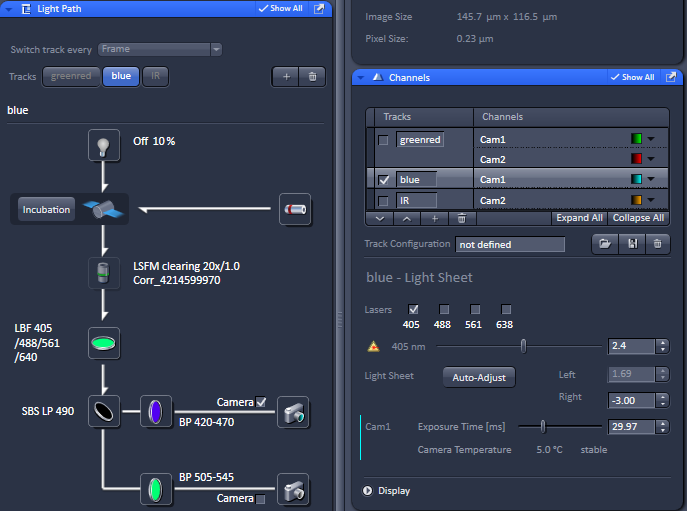

Camera 1 and Camera 2 need to be better aligned. These are XY images, green camera 1 and red camera 2.