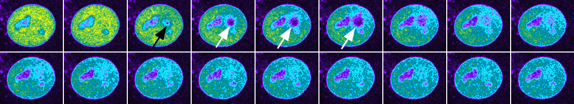
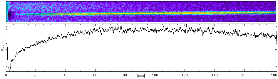
runavg8, med1x1, every 8th frame, 1st half of sequence, timescale explained below
20171219
Goal: to scan resonant mode fast while synchronized and simultaneous galvo photoactivation.
Conclusion: worked well
Test sample as per Young et al 2015 to induce DNA damage by localized exposure to UV light followed by recruitment of fluorescently labeled protein.
Microscope appears to be a mature, more sensitive, stable, and easy to use version of Zeiss 5 Live DuoScan used to FRAP podosomes (2007) and intended for later studies but switched to TIRF largely due to technical hurdles with the confocal. (For later: trythe same with spinning disk confocal.)
![]()
runavg8, med1x1, every 8th frame, 1st half of sequence, timescale explained below
Each frame 0.07 sec. XY 0.1 um/pixel. Meteadata as read from ND2 file by BioFormats here.
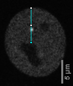
Kymogram location drawn on timpoint 669
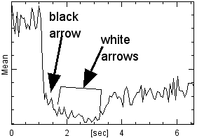
with kymogram and intensity curve on same timescale
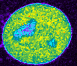
Overview with, from left to right,
original,
median filtered 1x1,
max pixel projection frames 105 to end threshholded and autowanded for intensity measurement through time,
running avg 8 through time.
Below are the intensity curves from the median and the runavg8 images.
![]()
Just the first 6 seconds measured onthe med1x1:
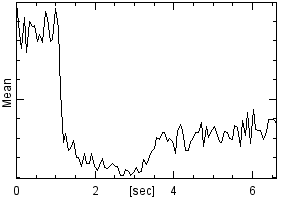
The dip in the curve shows a lot of bleaching during the photoactivation. Probably a briefer pulse of 405 nm laser would be enough to induce the photodamage and could improve kinetics of recruitment. The image below shows the bleach spot. These images are at 8 frames intervals after runavg8.