TNTs (tunneling nanotubes) before they were named?
The two pictures below from Am J Pathol. 1995 Jun;146(6):1508-20. Rojkind M1, Novikoff PM, Greenwel P, Rubin J, Rojas-Valencia L, de Carvalho AC, Stockert R, Spray D, Hertzberg EL, Wolkoff AW. Characterization and functional studies on rat liver fat-storing cell line and freshly isolated hepatocyte coculture system. PMID: 7778689 PMCID: PMC1870907 are confocal images that contain what appear to be tunneling nanotubes. "FSCs [fat storing cells] send out long projections and make contact with
hepatocytes that are some distance away." But it may have been too ambitious to say that the centriole had MTs radiating out the the point of contact. Also, in retrospect, I think we may be seeing background fluorescence as well as tubulin staining, so not convinced the projections are really lighting up due to MTs.
Twenty-five years later, at an ASCB/EMBO conference (Philadelphia 2017), I remembered these images and dug them up. Two years later I am posting them because they certainly appear to be TNTs. The orange arrows are added to point to likely TNTs.
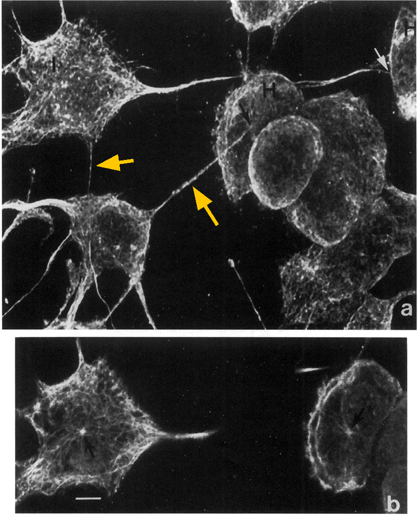
Figure 2. Confocal microscope images ofcocultures sbowing the localization oftubulin in cytoplasmic microtubules. a: Projection of31 serial optical sections of the entire depth ofFSCs (range of 12 to 18 t4 measured at cell center) and of hepatocytes (range of 20 to 31 4 measured at cell
center). The spatial relations ofFSCs (I) and hepatocytes (H) are evident. Note the long tubulin-positive projections ofFSCs (range of 8 to 14 ,u in
depth and 5 to 9 ,u in length) extending to the hepatocyte and contacting a small region ofthe hepatocyte surface (arrows). b: A 1-,u optical section
within the above projection of sections showing tubulin in a centriole (arrow) of a FSC (1) (same cell as upper left in a) and a hepatocyte (H)
(same cell as upper right in a). In the hepatocyte and FSC, elongated microtubules extendfrom the centriole to the cell surface; in the FSC, microtubules are also seen within its projections. Bar, 10 um.
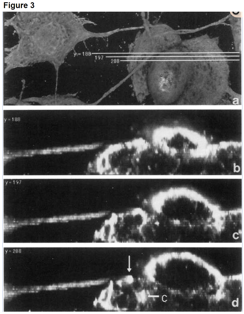
Figure 3. Confocal microscope images ofcocultures immunostainedfor tubulin. a: Volume of FSCs and hepatocytes rendered with Voxel View. b to d: y axis tews examined at three different points in the area of contact between a FSC extension and a hepatocyte. Cytoplasmic microtubules appear white; note the presence of microtubules beneath the area of contact (arrow). In (d), the area of contact is above a centriole region (C). Microtubules appear to extendfrom the centriole to the surface beneath the contact area. Bar, 10 um.
Technical details: fixation included glutaraldehyde which may have been necessary to preserve the delicate structures.
The image was on the journal cover too.

<back