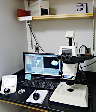
Leica dissection microscope with fluorescence
To sign up for microscope in advance, use Histopath Core reservations webpage on Labvantage LIMS. Leica M165 Fluorescent Stereo (dissecting) Scope
This microscope has grayscale brightfield and GFP and RFP flourescence.
If you want the details in 150 pages, Leica's PDF manual.
How to Turn On (follow step-by-step):
Methods for automatically taking multichannel images can be strung together.
The best way to save files is to group them within an experiment in Leica format. Each sample would be saved as a different experiment. This means you can take multiple pictures of each sample and only have to enter a file name once.
Images are easy to open in ImageJ and we have macros we can tweak to apply colors and automatically make figures in any format required. For instance, we can have ImageJ open every image in a directory and make montages of green, red, brightfield, and merged with a scalebar and saved with the correct filename. Automation saves a lot of time. Ask the Microscopy Core staff for help with this.
This microscope does not have a stage ideal for tiling, but tiling can be done.
If software crashes, data may be recoverable if you don't run the software first. The raw data may be in a Leica directory where they can be copied out before restarting the software.
Section to be written: fluorescence microscopy settings and how to take pictures in linear range for quantification.
Notes from day training with Leica: Two switches knobs on side of base change baffles and angles, clicks to be reproducible explain intensity max - change intensity otherwise Starting software: camera needs to be stopped to adjust microscope |
comments, questions, suggestions for this web page: Michael.Cammer@med.nyu.edu or mcammer@gmail.com