Bead Assay via Microscopy
Question: what is the concentration (or mass) of protein in a cell membrane?
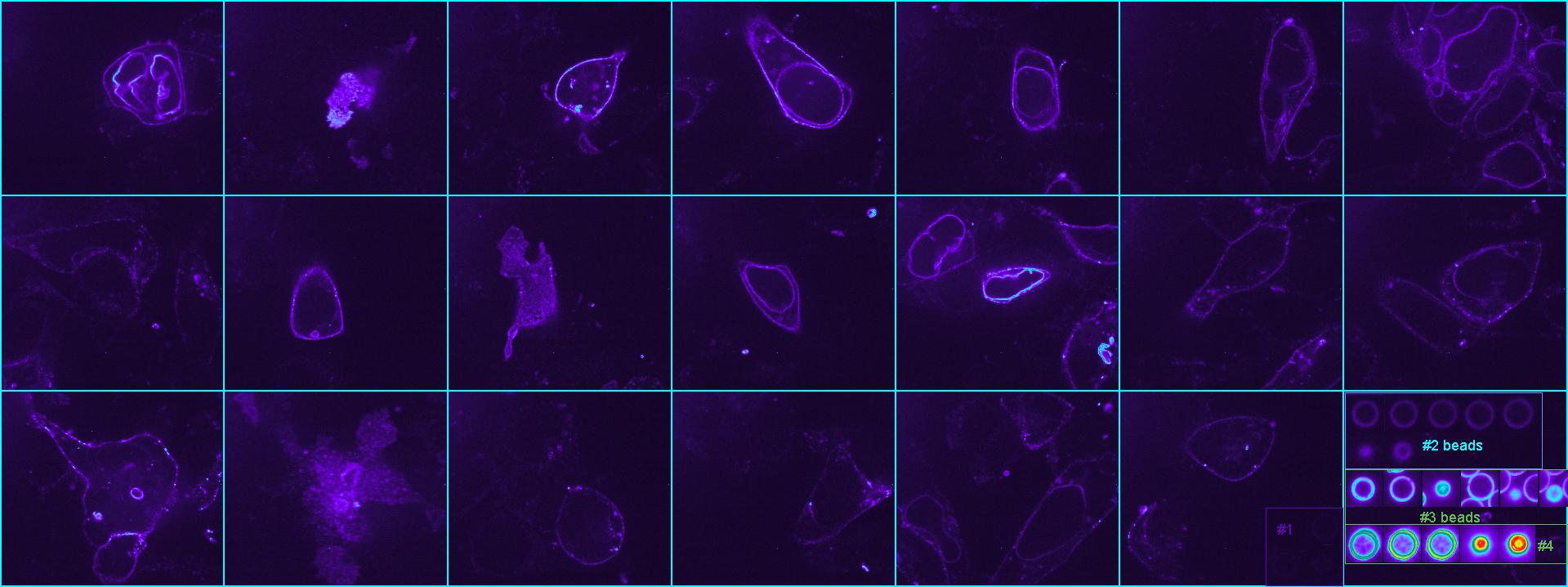
Using flow cytometry beads of known protein concentration imaged by spinning disc confocal with the cells the researchers intended to report the amount of protein in the membrane.
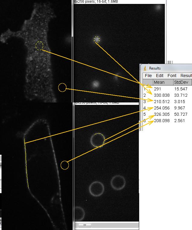
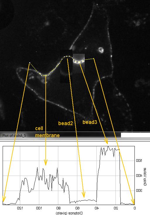
The goal was to compare the known mass and concentration standardized by flow cytometry with the intensity of the same protein on the membrane. The pictures below show the idea of how to do this.
However, the fluorescent probes used on the beads were different than the GFP expressed in the cell membranes, so the two really could not be directly compared. But to follow the method below, you may pretend that the protein and fluorophore on the bead is the same as in the real live cells.
Keep in mind the geometry of the imaging. Because the confocal optical section is thicker than the bead surface or the cell surface, we imaged at the equator of the bead and where the cell membrane was oriented vertically.
Bead standards # of molecules are:
beads0 0
beads1 3507
beads2 32348
beads3 196948
beads4 941222
25 Jan 2012
comments, questions, suggestions: Michael.Cammer@med.nyu.edu