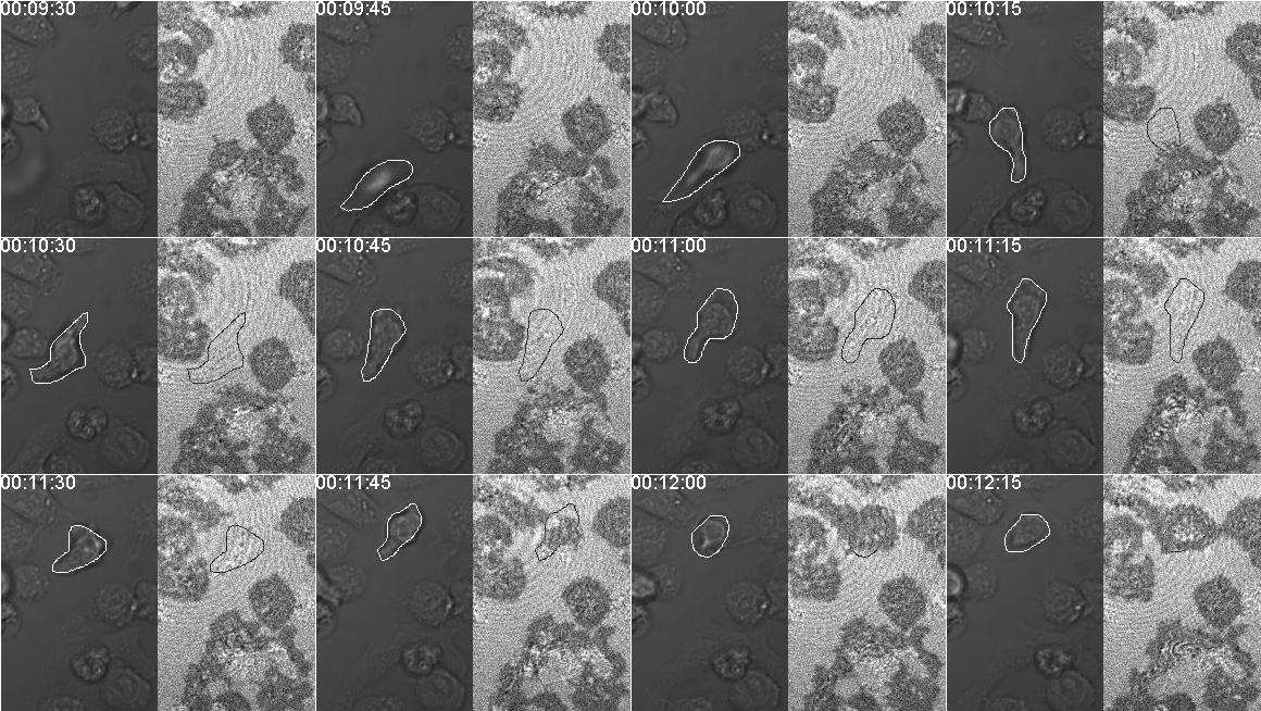
This page shows how IRM can be used to look at how well cells are attached to the substrate.
A few t cells are attached and spread to an ICAM-1 coated coverslip. (Each is approx 10 um in diameter.) A new one drops down from the media and begins crawling. It is attached enough to use the coverslip for traction for motility along the substrate, but it is attached by very little surface area so that in the IRM mode it is not visible about the specular background noise.
It then spreads and attaches to the substrate and is visible as a black spot. Look at timepoints 11:30 to 11:45. At 12:15, is appears to not be attached at the center where the may be a pocket with extruded material, perhaps an immunological synapse.
Imaged with a 63X N.A. 1.4 planapochromatic lens on a Zeiss 710. We probably used a 488 nm laser, but it may have been the 543 nm laser.
Circa 2012 with cells prepared by Hila Novack in Dustin lab. (Also posted at https://www.flickr.com/photos/mcammer/7726410962 and https://www.flickr.com/photos/mcammer/7726411530/.)
Here is a published example where frustrated phagocytosis was measured. https://www.nature.com/articles/ncb805/figures/8 from Myosin X is a downstream effector of PI(3)K during phagocytosis.

last updated 2022-02-09 20:12