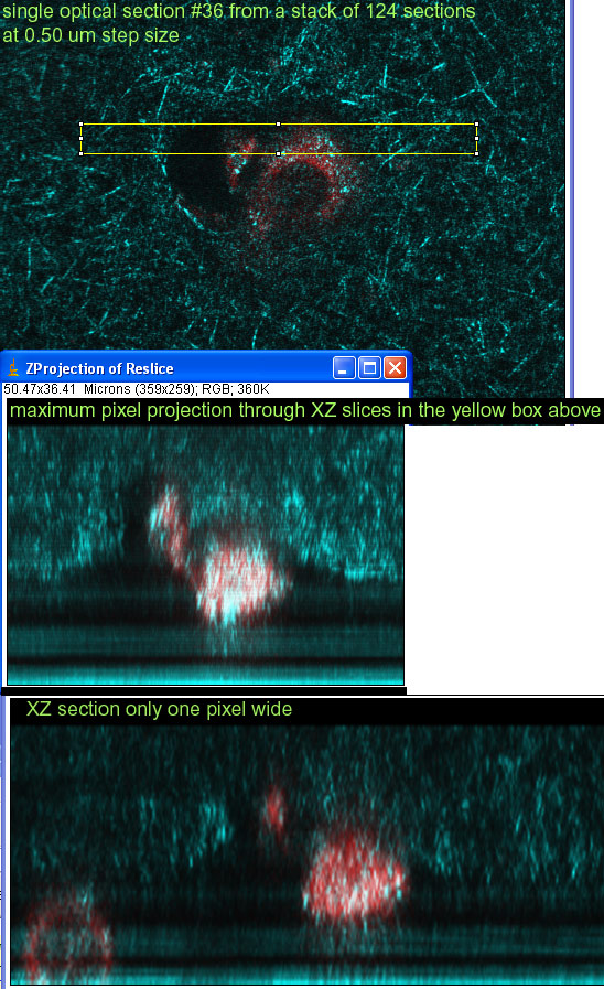
green: f-actin red: cell tracker
cells stained on 20040317 and imaged with BioRad Radiance confocal on 20040322
Single optical section through cell.
green: f-actin red: cell tracker
![]()
Cells plated on a coverslip below a layer of collagen do climb up into & through the collagen.
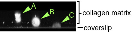
Coverslip at bottom.
Space of approx 11 um with media.
collagen matrix above.
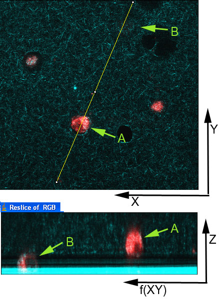
The image above is a single optical section through the collagen matrix.
The image below is a XZ section along the yellow line in the image above.
Cells were imaged with reflection at 488 nm and a red dye excited at 568 nm.
A good way to see both reflection and the tracer dye would be to stain the actin for excitation at 633 nm (e.g. Cy5 or Alexa 633).
A few more links to pics:
https://www.flickr.com/photos/mcammer/8805209354/ (Zeiss 710)
https://www.flickr.com/photos/mcammer/5871564798/ (BioRad Radiance)
https://www.flickr.com/photos/mcammer/5871008067/ (BioRad Radiance)
https://www.flickr.com/photos/mcammer/5871564888/ (BioRad Radiance)
(should add Bailly macro link)