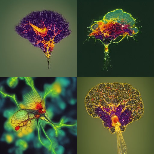
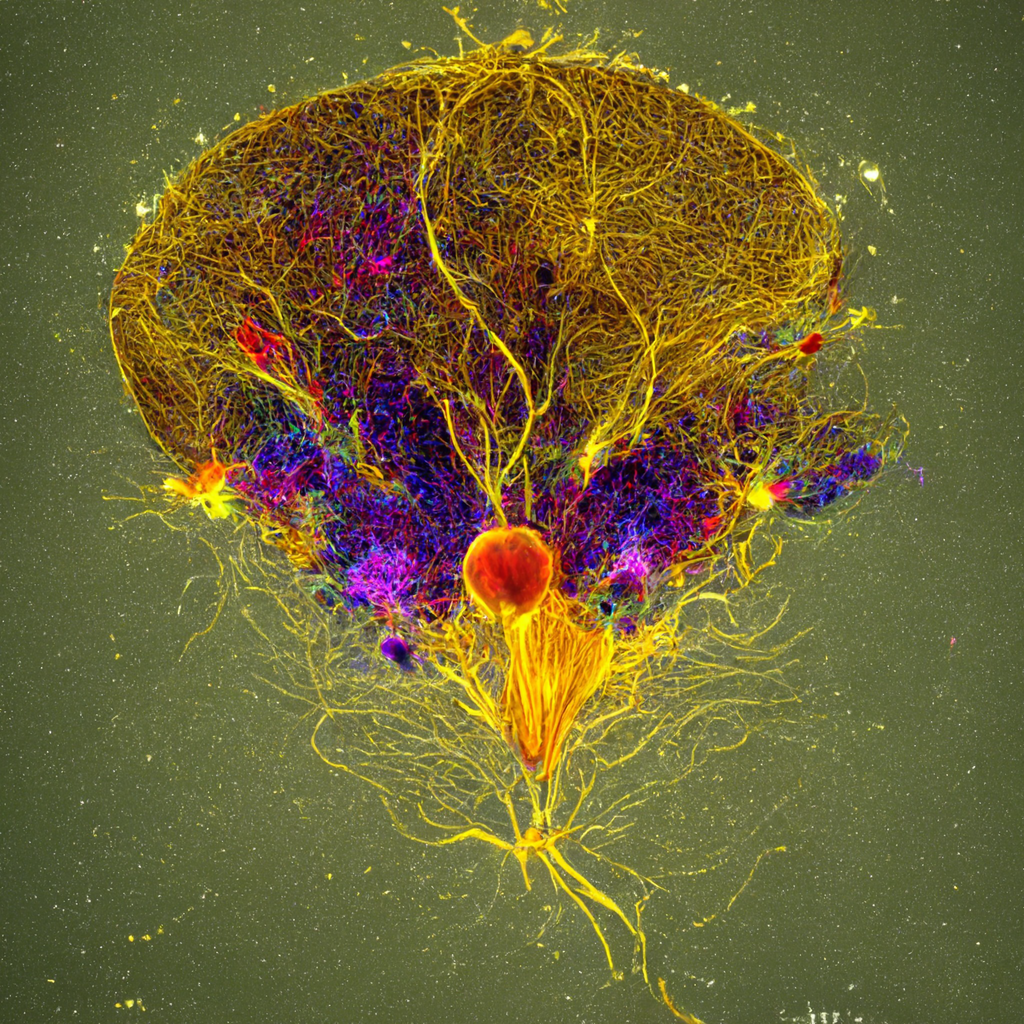
map of drosophila brain neurons microscopy, fill the field
I chose the lower right image as a seed for variations which follow.
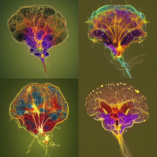
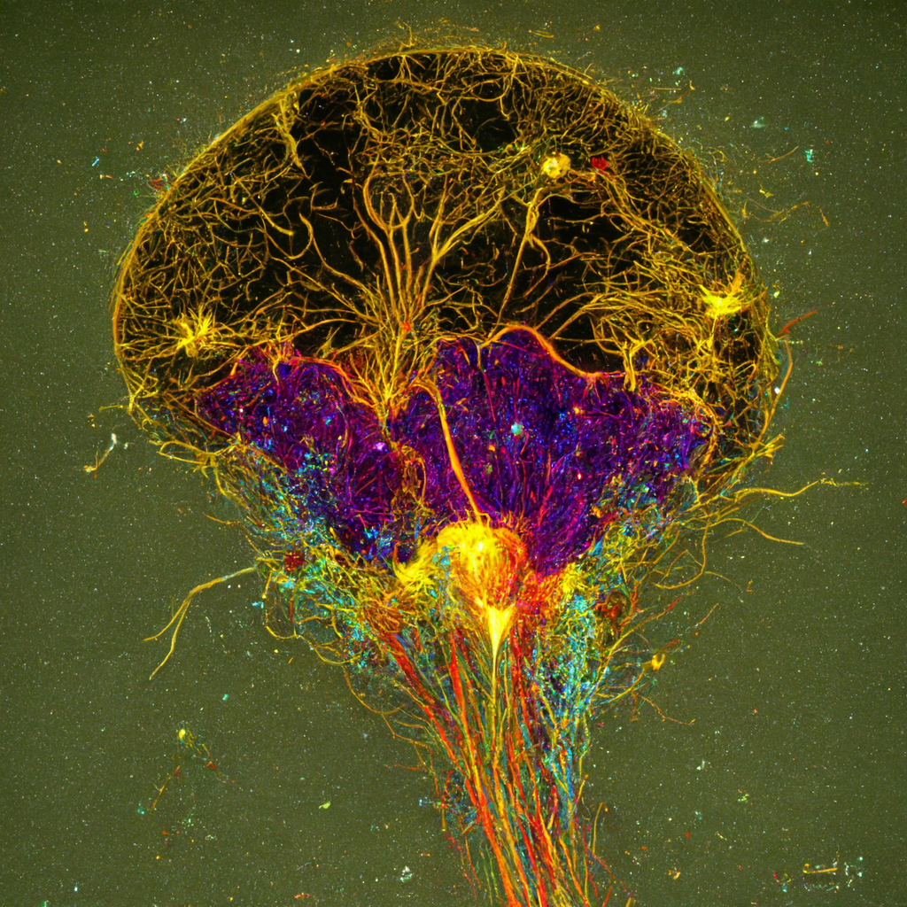
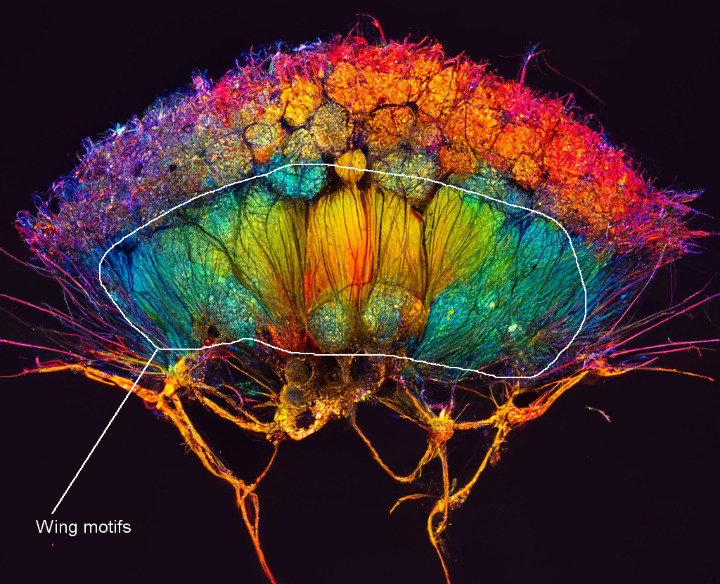
The following images are next generation of variations.
Clearly the AI doesn't have enough "understanding" of neuron distributions inside the brain; the tendrils look kind of like cooked spaghetti without but not clear oranization, directionality or destinations. But with more training, wow, this could generate amazing fake data. (AIs might also provide insight into organizations in the training sets not yet observed, so it may not all be bad.)
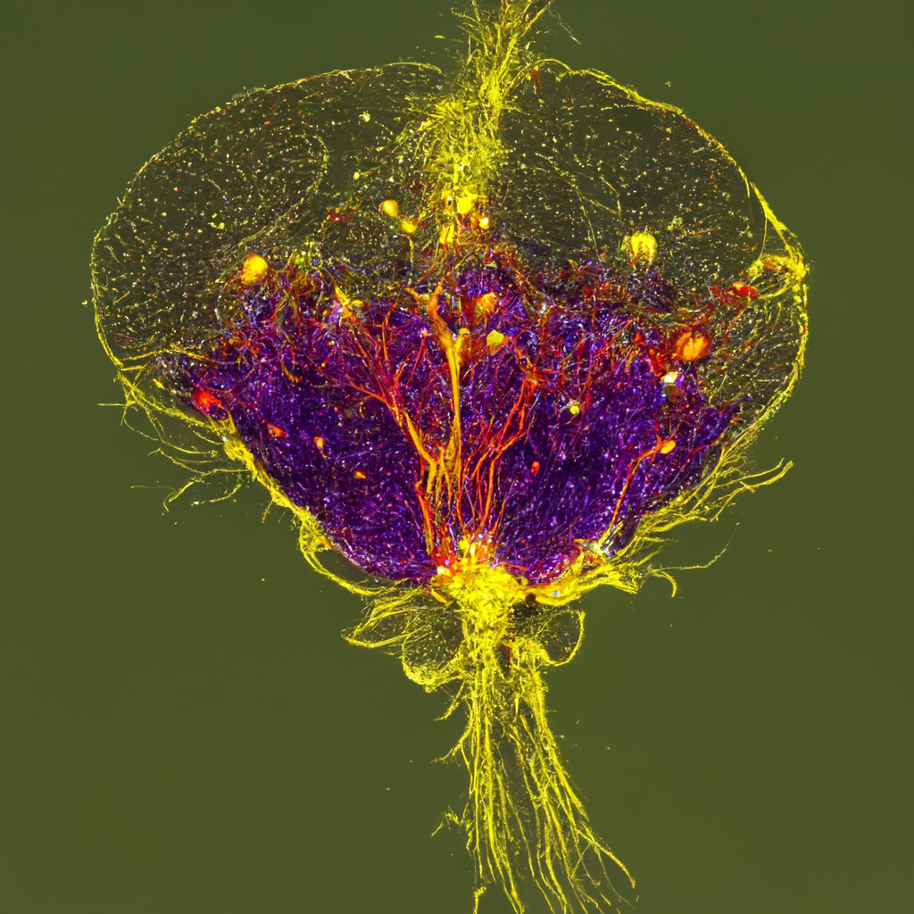
Somebody else in the Discord chat room saw what I was doing and chose to create variations of the lower left image of the first image set. I was not interested in this one initailly, but in a nod to validating group science, here it is.
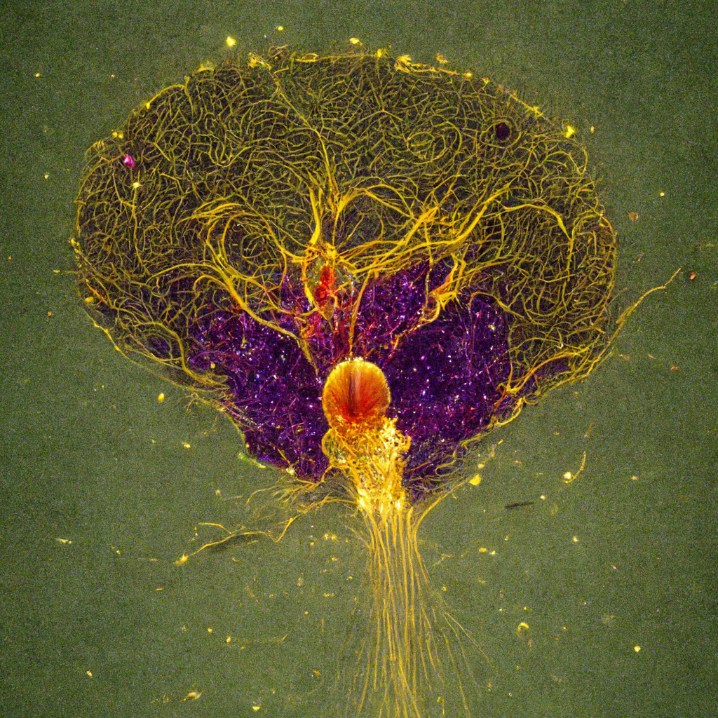
Upscaling the original one also yields additional information. Super resolution has never been so easy!
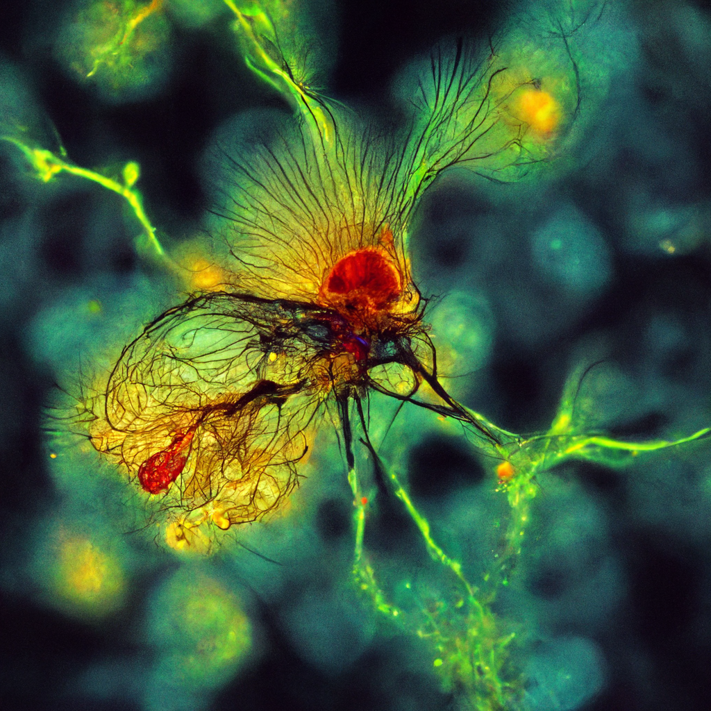
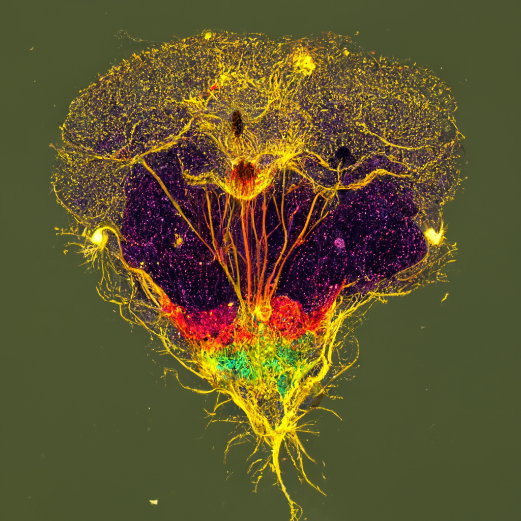
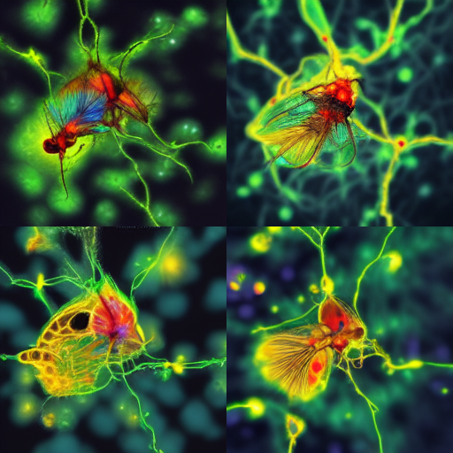
Another example of scrambled anatomy:
What if the variations were directed rather than random (or based on rules in the software we are unaware of). Fake data could get sophisticated.
Note that terms such as these were understood as essential properties of the terms I used:
bilateral symmetry and dorsal ventral , single bundle out and fraying with rough prep
The original commands were:
map of drosophila brain neurons microscopy, fill the field
The "fill the field" direction was ignored or not operated on. This is common to how the algorithm works. I looked at images being generated by other people and typically it would make compositions very central.
=========================
A few images of real fly brain based on the search:
https://www.google.com/search?q=map+drosophila+brain&rlz=1C1GCEU_enUS865US865&sxsrf=AJOqlzVosDkkk0xh_jsaRP95lCq0CmBtWg:1675699474140&source=lnms&tbm=isch&sa=X&ved=2ahUKEwjL35__ooH9AhUZL1kFHe0qBYkQ_AUoAXoECAEQAw&biw=1471&bih=1174&dpr=1
https://www.technologynetworks.com/neuroscience/articles/unlocking-the-secrets-of-brain-organization-in-the-fruit-fly-310488
https://www.janelia.org/project-team/flyem/blog/my-em-data-is-segmented-now-what
https://www.janelia.org/#&gid=1&pid=1
https://www.janelia.org/#&gid=1&pid=4
https://singularityhub.com/2021/12/28/the-biggest-brain-maps-ever-created-are-pushing-the-frontiers-of-neuroscience/
https://ai.googleblog.com/2020/01/releasing-drosophila-hemibrain.html
Note that AI was used heavily to segment the images and to generate network.
https://www.popularmechanics.com/technology/a30642613/google-3d-map-fly-brain/
A database of fly brain images labeled per gene:
https://gen1mcfo.janelia.org/cgi-bin/view_gen1mcfo_imagery.cgi?line=R10A09
===========================
At this point, I decided to return to the cell culture terms. More -->
Later attempts (2023-02-05) in Dall-e yielded results so far from anything resembling fly and so aesthetically uninteresting that not posting here.
first written 2022-10-24 21:45
mcammer@gmail.com