Point of this webpage is a practical explanation of confocal pinhole.
Conclusions regarding quality of images in XY axes:
Pinhole at 1.5 Airy units result in images noticeably less resolution than pinhole at 1.0.
Closing pinhole, such as to 0.7 Airy units, is noticeably better than 1.0. (Not discussed, how this may increase bleaching rate or noise.)
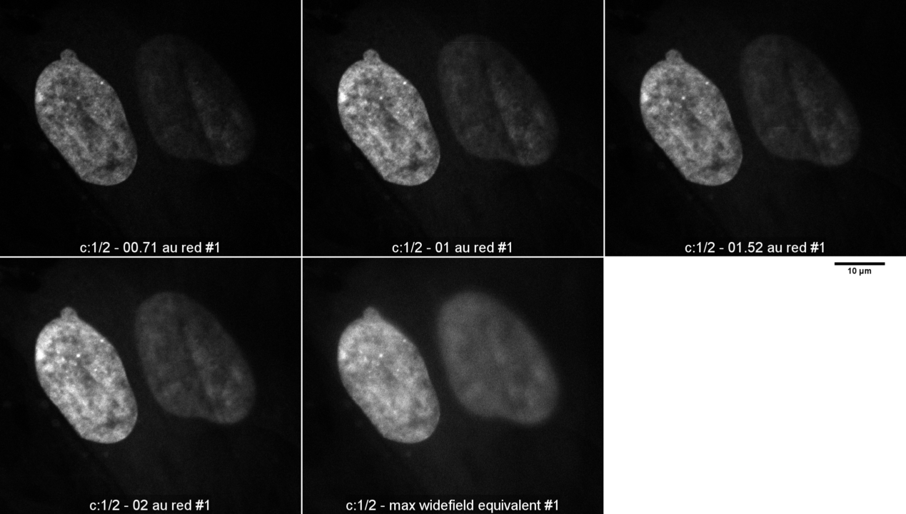
Sample is mammalian cell culture on #1.5 coverslip stained for smooth muscle actin with red fluorescent probe (Alexa 555 or 568) and mounted in Prolong Gold. Imaged with 63X N.A. 1.4 planapochromat DIC on Zeiss 880 with GaAsP detector.
Images were taken with pinhole, as calculated by Zeiss Zen Black software, at 0.71, 1.0, 1.52, 2.0, and maximum opened. Laser was attenuated between pinhole settings. Gain, scan speed, averaging kept constant for all pictures. Original images 1024 x 1024 pixels oversampled at each pixel 0.07 um. Other metadata here.
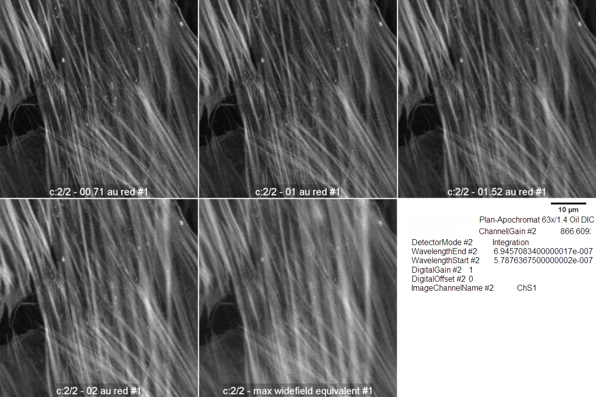
Images below shown at 0.5X original scale.
These images had gamma 0.6 applied to better see details in the lower intensity areas.
My assessment: by eye, the 0.71 Airy unit image looks a bit better than the 1 A.U. The 1.52 A.U. and the 2 A.U. image look the same, but the 1 A.U. image is noticeably sharper.
The maximum pinhole image is fuzzy.
Quantitative analysis using FFT, which is shown at the bottom of this page, agrees with my visual perception.
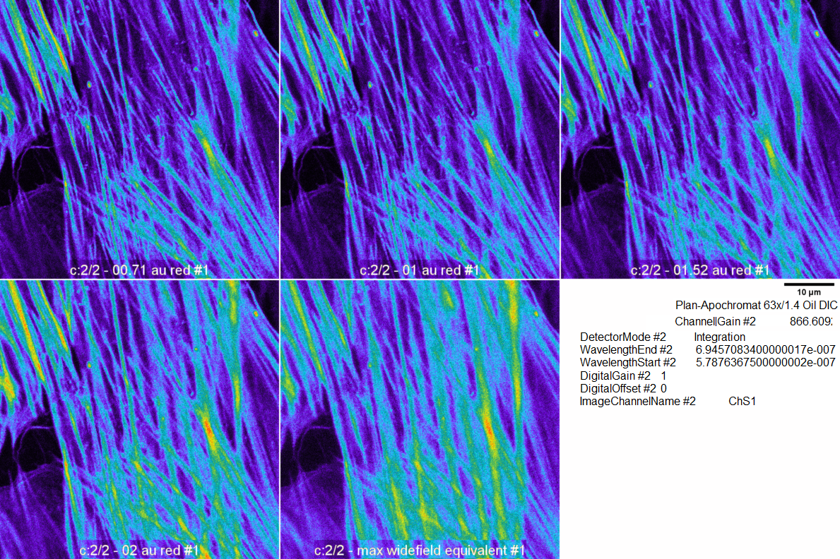
These images are the original linear scale but with a color look up table applied.
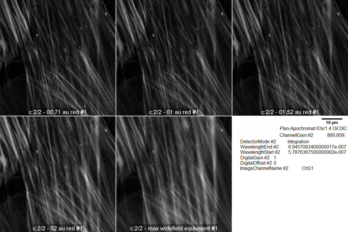
These images are the original raw data with intensity displayed linearly.
For each pinhole setting, the ImageJ FFT command was executed on the whole original image. The larger the power spectrum, the more high frequency (i.e. resolution).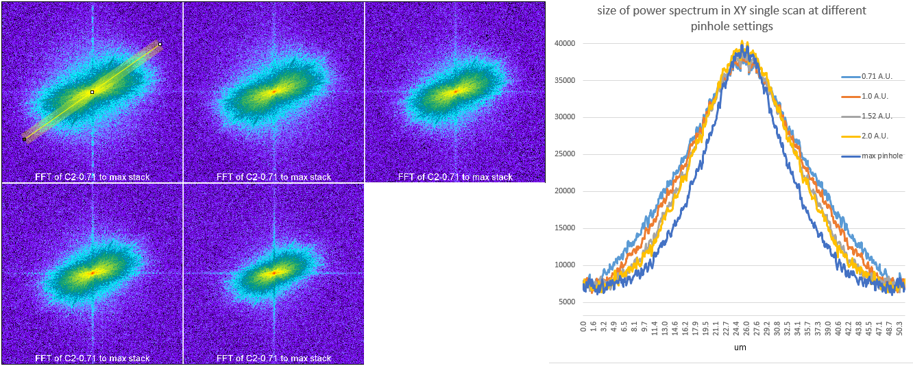
Also, the sample was stained with dapi, but I wasn't as careful to match intensities.
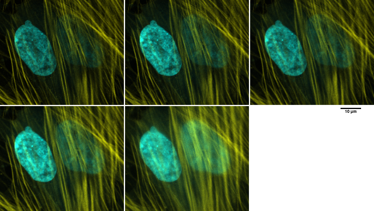
(The occasional horizontal lines were because the AOTF was acting up. Has been been fixed.)
<Table Of Contents
raw data at R:\MicroscopyCore\mcammer\equipment profiles\880\20190625 smoothmuscleactin\2