Zeiss 710 MP microscope
A summary of first tests with the inverter which shows intensity loss when using the multi-photon: summary01.pdf
This loss of intensity does not appear to happen with the normal confocal operation
(to be confirmed with a bead test), which means that the loss is on the collection side, not the excitation side, due to less collection of scattered emission.
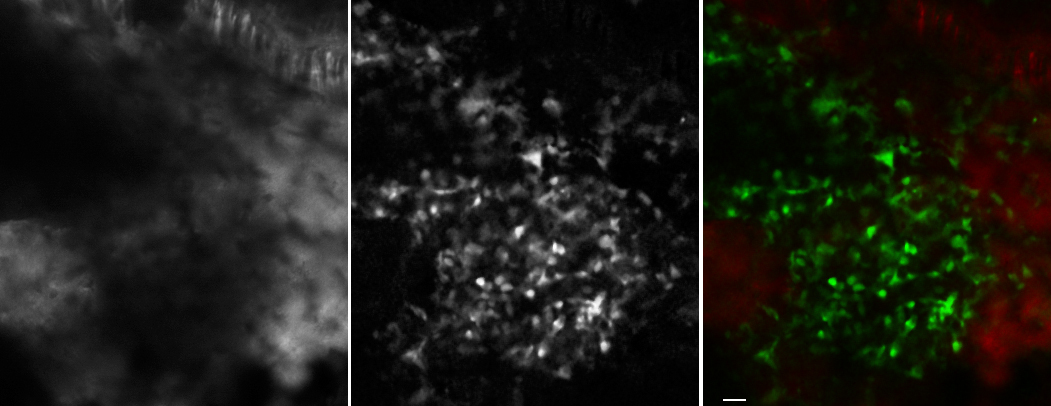
Z series collected at 512 X 512 with 20X water N.A. 1.00 objective. No zoom was used. We think each pixel is 0.42 X 0.42 um. The Z step size 0.84 um. The following picture is an average projection of the stack after 1X1 median filtering of slices 75 though 85 (9 um) of the 188 slice stack (158 um deep total). Scalebar 10 um. Tissue is mouse skull in vivo prepared by Jiyun Kim. Bone marrow niche in a cx3cr1 gfp knock in heterozygous mouse. Red is SHG signal from surrounding bone.
A movie of the Z series from deep to surface: mouse_skull.avi
The Z axis resolution is not great, and, a caveat with normal confocal is that we do not kow whether blue, green, and red are all parfocal in the Z axis.
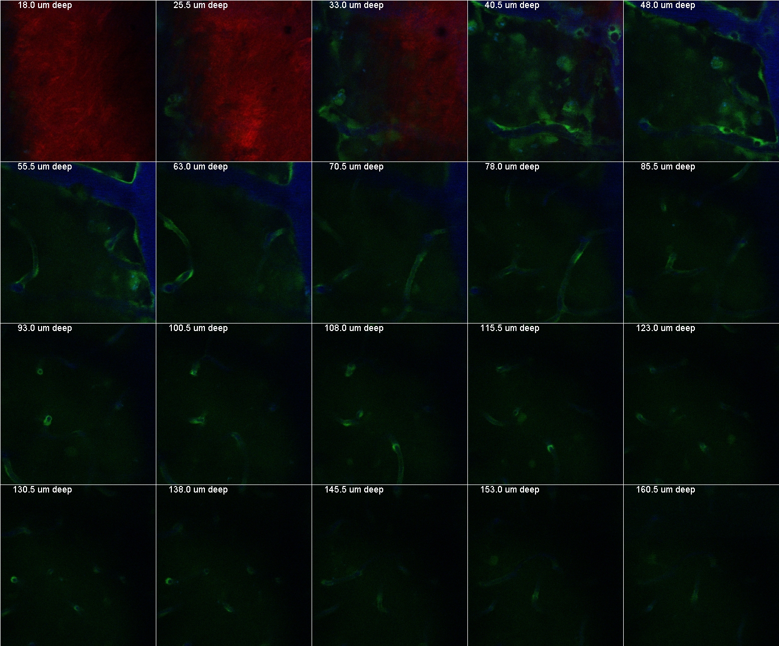
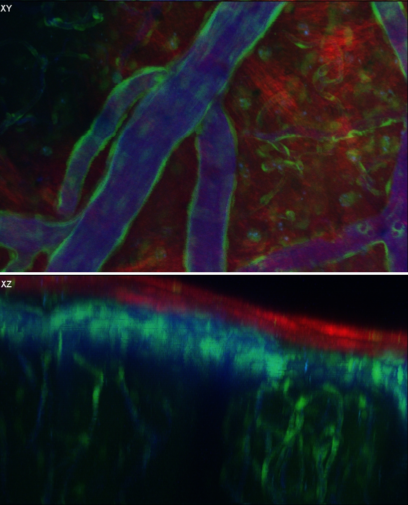
The following in vivo images were collected by PJ and then prepared for this webpage by MC using ImageJ.
Individual Z sections:
Maximum pixel projection from above and side (with depth cueing):
A benefit of the 20X objective is that it scans 4X the area in a single field as the 40X water objective at the same resolution. Because it has the same numerical aperture as the 40X water, at 1024X1024 pixels the image is the same resolution as tiling four separate scans with the 40X at 512X512.
comments, questions, suggestions: Michael.Cammer@med.nyu.edu
Link to main lab pages: http://saturn.med.nyu.edu/research/mp/dustinlab/