Zeiss 710 MP microscope
Beads tests for Z axis resolution
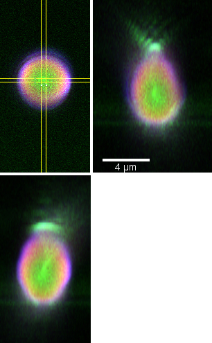 |
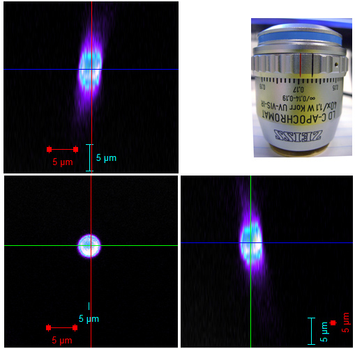
Standard confocal
4um Invitrogen Tetraspeck beads dried onto a 1.5 coverslip and then mounted in water.
Imaged Oct 2010 with 63X oil objective, pihole1 airy unit, XY 0.006 um/pixel, Z step 0.18 um, scan speed 6.30 usec at 256 X 256, average 4 frames. Laser lines 488, 543, 647 nm & 3 channels collected simultaneously and set for no spillover from one channel into another.
From the equator down to below coverslip looks good. |
Note the reduced resolution in the Z axis.
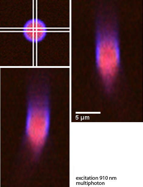
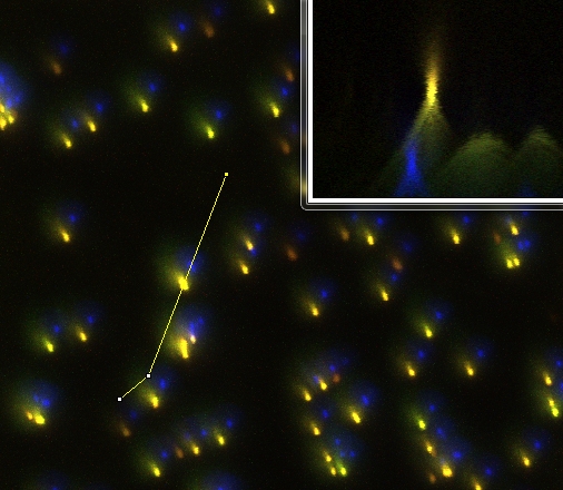
Approximately 3 X 3 X 2 mm plug of ground fatty muscle (a.k.a. pork sausage with n-propyl-gallate as preservative) mixed with 20 ul of 4.0 um Tetraspeck beads pushed to the bottom of a Lab-Tek coverglass bottom chamber. Excitation at 910 nm. Focused deep into material and then adjusted DeepSee motor (position approx 56) to optimum brightness.
Also tried imaging with the 25X N.A. 0.8 objective and only got approx 50 um deep, but not in exact same area of tissue.
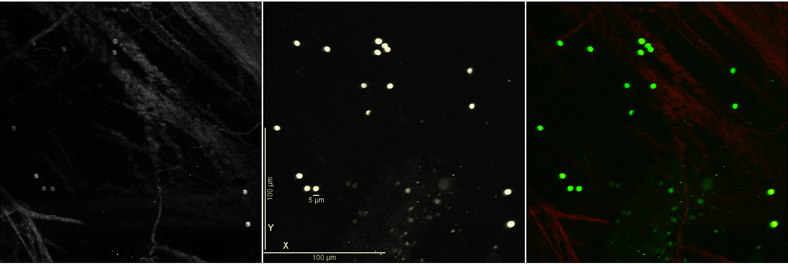
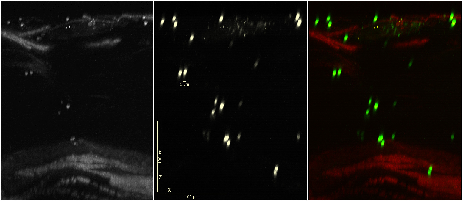
Approx 50 um in with zoom:
All the Z series above look slanted, off axis. When evaluating a 20X N.A. 1.0 longer working distance objective we noticed that not only was the slant bad, but red light which focused at a very different plane than the blue/green was offset. (As shown here, RGB corresponds to 422-470 nm; 518-616 nm; and 645-735 nm.)
Back
comments, questions, suggestions: Michael.Cammer@med.nyu.edu
Link to main lab pages: http://saturn.med.nyu.edu/research/mp/dustinlab/